
Page 677 - Atlas of Histology with Functional Correlations
P. 677
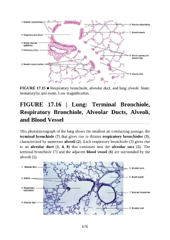
FIGURE 17.15 ■ Respiratory bronchiole, alveolar duct, and lung alveoli. Stain:
               hematoxylin and eosin. Low magnification.


               FIGURE  17.16  |  Lung:  Terminal  Bronchiole,

               Respiratory  Bronchiole,  Alveolar  Ducts,  Alveoli,


               and Blood Vessel


               This photomicrograph of the lung shows the smallest air-conducting passage, the
               terminal bronchiole (7) that gives rise to thinner respiratory bronchioles (3),
               characterized by numerous alveoli (2). Each respiratory bronchiole (3) gives rise

               to  an  alveolar  duct  (1,  4,  8)  that  continues  into  the  alveolar  sacs  (5).  The
               terminal bronchiole (7) and the adjacent blood vessel (6) are surrounded by the
               alveoli (2).


                                                          676