
Page 671 - Atlas of Histology with Functional Correlations
P. 671
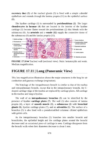
excretory duct (5)  of  the  tracheal  glands  (3)  is  lined  with  a  simple  cuboidal

               epithelium and extends through the lamina propria (11) to the epithelial surface
               (8).

                   The hyaline cartilage (1) is surrounded by perichondrium (2). The larger
               chondrocytes  in  lacunae  (4)  that  are  located  in  the  interior  of  the  hyaline
               cartilage (1) become flatter toward the perichondrium (2) that blends with the

               submucosa (6). An arteriole and a venule (12) supply the connective tissue of
               the submucosa (6) and the lamina propria (11).


               FIGURE 17.10 ■ Tracheal wall (sectional view). Stain: hematoxylin and eosin.
               Medium magnification.


               FIGURE 17.11 | Lung (Panoramic View)


               This low-magnification illustration shows the major structures in the lung for air

               conduction and gaseous exchange (respiration).

                   The histology of the intrapulmonary bronchi is similar to that of the trachea
               and extrapulmonary bronchi, except that in the intrapulmonary bronchi, the C-
               shaped cartilage rings of the trachea are replaced by cartilage plates. All cartilage
               in the trachea and lung is hyaline.

                   The  wall  of  an  intrapulmonary  bronchus  (5)  can  be  identified  by  the

               presence  of  hyaline  cartilage plates (7).  The  wall  (5)  also  consists  of  lamina
               propria  (4),  a  layer  of  smooth  muscle  (3),  a  submucosa  (2)  with  bronchial
               glands (6), hyaline cartilage plates (7), and an adventitia (1). The surface of a

               bronchus (5) is also lined with a pseudostratified columnar ciliated epithelium
               with goblet cells.

                   As  the  intrapulmonary  bronchus  (5)  branches  into  smaller  bronchi  and
               bronchioles,  the  epithelial  height  and  the  cartilage  plates  around  the  bronchi
               decrease until an occasional piece of cartilage is seen. Cartilage disappears from

               the bronchi walls when their diameters decrease to about 1 mm.


                                                          670