
Page 669 - Atlas of Histology with Functional Correlations
P. 669
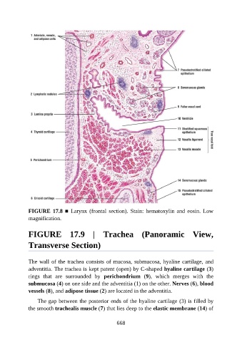
FIGURE 17.8 ■  Larynx  (frontal  section).  Stain:  hematoxylin  and  eosin.  Low
               magnification.


               FIGURE  17.9  |  Trachea  (Panoramic  View,

               Transverse Section)


               The wall of the trachea consists of mucosa, submucosa, hyaline cartilage, and
               adventitia. The trachea is kept patent (open) by C-shaped hyaline cartilage (3)
               rings  that  are  surrounded  by  perichondrium  (9),  which  merges  with  the

               submucosa (4) on one side and the adventitia (1) on the other. Nerves (6), blood
               vessels (8), and adipose tissue (2) are located in the adventitia.

                   The gap between the posterior ends of the hyaline cartilage (3) is filled by
               the smooth trachealis muscle (7) that lies deep to the elastic membrane (14) of


                                                          668