
Page 662 - Atlas of Histology with Functional Correlations
P. 662
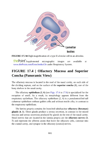
FIGURE 17.3 ■ High magnification of a type II alveolar cell in an alveolus.


                                Supplemental       micrographic       images     are    available     at
                 www.thePoint.com/Eroschenko13e under Respiratory System.


               FIGURE  17.4  |  Olfactory  Mucosa  and  Superior

               Concha (Panoramic View)


               The olfactory mucosa is located in the roof of the nasal cavity, on each side of
               the dividing septum, and on the surface of the superior concha (1), one of the
               bony shelves in the nasal cavity.

                   The olfactory epithelium (2, 6) (see Figs. 17.4 to 17.6) is specialized for the

               reception  of  smell.  As  a  result,  its  morphology  appears  different  from  the
               respiratory epithelium. The olfactory epithelium (2, 6) is a pseudostratified tall
               columnar epithelium without goblet cells and without motile cilia, in contrast to
               the respiratory epithelium.

                   The lamina propria contains the branched tubuloacinar olfactory (Bowman)

               glands (4, 5). These glands produce a serous secretion, in contrast to the mixed
               mucous and serous secretions produced by glands in the rest of the nasal cavity.
               Small nerves that are located in the lamina propria are the olfactory nerves (3,

               7) and represent the afferent axons that leave the olfactory cells, continue into
               the cranial cavity, and synapse in the olfactory (cranial) nerves.


                                                          661