
Page 747 - Atlas of Histology with Functional Correlations
P. 747
as Herring bodies. When needed, hormones from the neurohypophysis are
released and enter the adjacent fenestrated capillaries in the pars nervosa. Thus,
the neurohypophysis functions as a storage site for neuroendocrine secretions
that were synthesized in and transported from the supraoptic and paraventricular
nuclei of the hypothalamus.
Supplemental micrographic images are available at
www.thePoint.com/Eroschenko13e under Endocrine System.
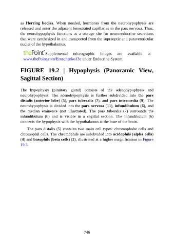
FIGURE 19.2 | Hypophysis (Panoramic View,
Sagittal Section)
The hypophysis (pituitary gland) consists of the adenohypophysis and
neurohypophysis. The adenohypophysis is further subdivided into the pars
distalis (anterior lobe) (5), pars tuberalis (7), and pars intermedia (9). The
neurohypophysis is divided into the pars nervosa (11), infundibulum (6), and
the median eminence (not illustrated). The pars tuberalis (7) surrounds the
infundibulum (6) and is visible in a sagittal section. The infundibulum (6)
connects the hypophysis with the hypothalamus at the base of the brain.
The pars distalis (5) contains two main cell types: chromophobe cells and
chromophil cells. The chromophils are subdivided into acidophils (alpha cells)
(4) and basophils (beta cells) (2), illustrated at a higher magnification in Figure
19.3.
746