
Page 751 - Atlas of Histology with Functional Correlations
P. 751
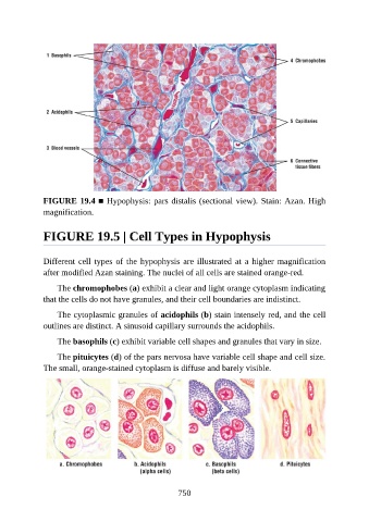
FIGURE 19.4 ■ Hypophysis: pars distalis (sectional view). Stain: Azan. High
               magnification.


               FIGURE 19.5 | Cell Types in Hypophysis


               Different cell types of the hypophysis are illustrated at a higher magnification
               after modified Azan staining. The nuclei of all cells are stained orange-red.

                   The chromophobes (a) exhibit a clear and light orange cytoplasm indicating

               that the cells do not have granules, and their cell boundaries are indistinct.

                   The cytoplasmic granules of acidophils (b) stain intensely red, and the cell
               outlines are distinct. A sinusoid capillary surrounds the acidophils.

                   The basophils (c) exhibit variable cell shapes and granules that vary in size.

                   The pituicytes (d) of the pars nervosa have variable cell shape and cell size.

               The small, orange-stained cytoplasm is diffuse and barely visible.


                                                          750