
Page 748 - Atlas of Histology with Functional Correlations
P. 748
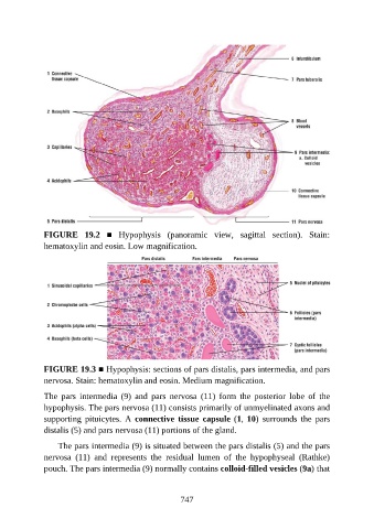
FIGURE  19.2  ■  Hypophysis  (panoramic  view,  sagittal  section).  Stain:
               hematoxylin and eosin. Low magnification.


               FIGURE 19.3 ■ Hypophysis: sections of pars distalis, pars intermedia, and pars
               nervosa. Stain: hematoxylin and eosin. Medium magnification.

               The  pars  intermedia  (9)  and  pars  nervosa  (11)  form  the  posterior  lobe  of  the
               hypophysis. The pars nervosa (11) consists primarily of unmyelinated axons and

               supporting  pituicytes.  A  connective  tissue  capsule (1, 10)  surrounds  the  pars
               distalis (5) and pars nervosa (11) portions of the gland.

                   The pars intermedia (9) is situated between the pars distalis (5) and the pars
               nervosa  (11)  and  represents  the  residual  lumen  of  the  hypophyseal  (Rathke)
               pouch. The pars intermedia (9) normally contains colloid-filled vesicles (9a) that


                                                          747