
Page 752 - Atlas of Histology with Functional Correlations
P. 752
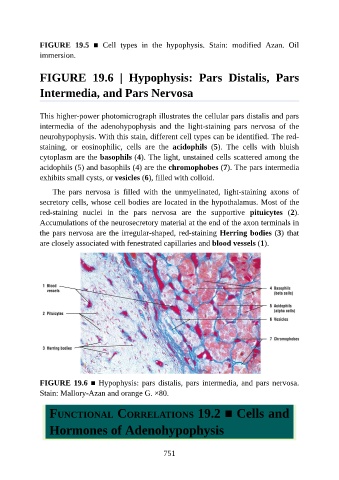
FIGURE  19.5  ■  Cell  types  in  the  hypophysis.  Stain:  modified  Azan.  Oil

               immersion.


               FIGURE  19.6  |  Hypophysis:  Pars  Distalis,  Pars


               Intermedia, and Pars Nervosa


               This higher-power photomicrograph illustrates the cellular pars distalis and pars
               intermedia  of  the  adenohypophysis  and  the  light-staining  pars  nervosa  of  the
               neurohypophysis. With this stain, different cell types can be identified. The red-

               staining,  or  eosinophilic,  cells  are  the  acidophils  (5).  The  cells  with  bluish
               cytoplasm are the basophils (4). The light, unstained cells scattered among the
               acidophils (5) and basophils (4) are the chromophobes (7). The pars intermedia
               exhibits small cysts, or vesicles (6), filled with colloid.

                   The  pars  nervosa  is  filled  with  the  unmyelinated,  light-staining  axons  of

               secretory cells, whose cell bodies are located in the hypothalamus. Most of the
               red-staining  nuclei  in  the  pars  nervosa  are  the  supportive  pituicytes  (2).
               Accumulations of the neurosecretory material at the end of the axon terminals in
               the pars nervosa are the irregular-shaped, red-staining Herring bodies (3) that

               are closely associated with fenestrated capillaries and blood vessels (1).


               FIGURE 19.6 ■ Hypophysis: pars distalis, pars intermedia, and pars nervosa.

               Stain: Mallory-Azan and orange G. ×80.


                  FUNCTIONAL CORRELATIONS 19.2 ■ Cells and

                  Hormones of Adenohypophysis


                                                          751