
Page 775 - Atlas of Histology with Functional Correlations
P. 775
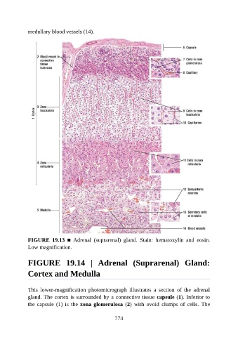
medullary blood vessels (14).


               FIGURE  19.13  ■  Adrenal  (suprarenal)  gland.  Stain:  hematoxylin  and  eosin.
               Low magnification.


               FIGURE  19.14  |  Adrenal  (Suprarenal)  Gland:

               Cortex and Medulla


               This  lower-magnification  photomicrograph  illustrates  a  section  of  the  adrenal
               gland. The cortex is surrounded by a connective tissue capsule (1). Inferior to
               the  capsule  (1)  is  the  zona glomerulosa  (2)  with  ovoid  clumps  of  cells.  The


                                                          774