
Page 776 - Atlas of Histology with Functional Correlations
P. 776
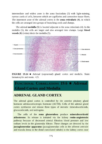
intermediate  and  widest  zone  is  the  zona  fasciculata  (3)  with  light-staining,

               narrow cords of cells, between which are capillaries and connective tissue fibers.
               The innermost zone of the adrenal cortex is the zona reticularis (4), in which
               the cells are arranged into groups of branching cords and clumps.

                   The adrenal medulla (5) is located adjacent to the zona reticularis (4). In the
               medulla  (5),  the  cells  are  larger  and  also  arranged  into  clumps.  Large  blood

               vessels (6) (veins) drain the medulla (5).


               FIGURE  19.14  ■  Adrenal  (suprarenal)  gland:  cortex  and  medulla.  Stain:
               hematoxylin and eosin. ×25.


                  FUNCTIONAL  CORRELATIONS  19.6  ■  Adrenal


                  Gland Cortex and Medulla


                 ADRENAL GLAND CORTEX


                 The  adrenal  gland  cortex  is  controlled  by  the  anterior  pituitary  gland
                 hormone  adrenocorticotropic  hormone  (ACTH).  Cells  of  the  adrenal  gland

                 cortex  synthesize  and  release  three  types  of  steroids:  mineralocorticoids,
                 glucocorticoids, and androgens.

                     The  cells  of  the  zona  glomerulosa  produce  mineralocorticoid
                 aldosterone.  Its  release  is  initiated  via  the  kidney  renin–angiotensin
                 pathway  because  of  decreased  arterial  filtration  blood  pressure  and  low

                 sodium levels in the glomerular filtrate. These changes are detected by the
                 juxtaglomerular apparatus (juxtaglomerular cells in the afferent arteriole
                 and macula densa in the distal convoluted tubule) in the kidney cortex near


                                                          775