
Page 772 - Atlas of Histology with Functional Correlations
P. 772
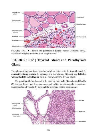
FIGURE  19.11  ■  Thyroid  and  parathyroid  glands:  canine  (sectional  view).
               Stain: hematoxylin and eosin. Low magnification.


               FIGURE 19.12 | Thyroid Gland and Parathyroid

               Gland


               This photomicrograph shows parathyroid gland adjacent to the thyroid gland. A
               connective tissue septum (3) separates the two glands. Different size follicles
               with colloid (1) and follicular cells (2) characterize the thyroid gland.

                   The parathyroid gland contains the smaller chief cells (4) and oxyphil cells
               (5)  that  are  larger  and  less  numerous  and  exhibit  an  eosinophilic  cytoplasm.

               Numerous blood vessels (6) surround the secretory cells in both organs.


                                                          771