
Page 768 - Atlas of Histology with Functional Correlations
P. 768
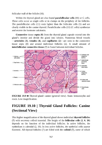
follicular wall of the follicle (10).

                   Within the thyroid gland are also found parafollicular cells (11) or C cells.

               These cells occur as single cells or in clumps on the periphery of the follicles.
               The  parafollicular  cells  (11)  stain  lighter  than  the  follicular  cells  (5)  and  are
               clearly visible in the canine thyroid. Parafollicular cells (11) (C cells) synthesize
               and secrete the hormone calcitonin.

                   Connective tissue septa (8) from the thyroid gland capsule extend into the

               gland’s  interior  and  divide  the  gland  into  lobules.  Numerous  blood  vessels
               —arterioles (3), venules (4),  and  capillaries  (2)—are  seen  in  the  connective
               tissue  septa  (8)  and  around  individual  follicles  (2).  A  small  amount  of

               interfollicular connective tissue (7) is found between individual follicles.


               FIGURE 19.9 ■ Thyroid gland: canine (general view). Stain: hematoxylin and
               eosin. Low magnification.


               FIGURE 19.10 | Thyroid Gland Follicles: Canine

               (Sectional View)


               This higher magnification of the thyroid gland shows individual thyroid follicles

               (7) with secretory colloid material. The height of the follicular cells (2, 6, 10)
               depends  on  the  function  of  the  individual  follicles.  In  active  follicles,  the
               epithelium is cuboidal (2, 10). In less active follicles, the epithelial cells appear

               flattened. All thyroid follicles (7) are filled with the colloid (7), some of which


                                                          767