
Page 767 - Atlas of Histology with Functional Correlations
P. 767
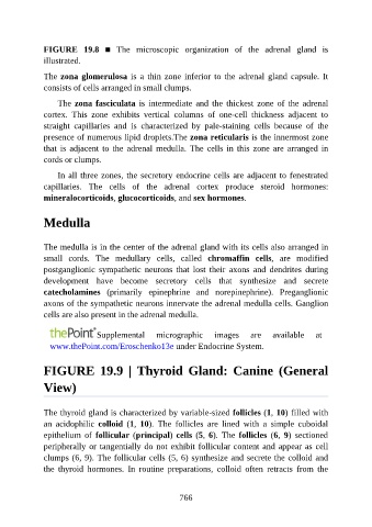
FIGURE  19.8  ■  The  microscopic  organization  of  the  adrenal  gland  is

               illustrated.

               The zona glomerulosa is a thin zone inferior to the adrenal gland capsule. It
               consists of cells arranged in small clumps.

                   The zona fasciculata  is  intermediate  and  the  thickest  zone  of  the  adrenal
               cortex.  This  zone  exhibits  vertical  columns  of  one-cell  thickness  adjacent  to
               straight  capillaries  and  is  characterized  by  pale-staining  cells  because  of  the

               presence of numerous lipid droplets.The zona reticularis is the innermost zone
               that  is  adjacent  to  the  adrenal  medulla.  The  cells  in  this  zone  are  arranged  in
               cords or clumps.

                   In all three zones, the secretory endocrine cells are adjacent to fenestrated

               capillaries.  The  cells  of  the  adrenal  cortex  produce  steroid  hormones:
               mineralocorticoids, glucocorticoids, and sex hormones.


               Medulla


               The medulla is in the center of the adrenal gland with its cells also arranged in
               small  cords.  The  medullary  cells,  called  chromaffin  cells,  are  modified
               postganglionic  sympathetic  neurons  that  lost  their  axons  and  dendrites  during
               development  have  become  secretory  cells  that  synthesize  and  secrete

               catecholamines  (primarily  epinephrine  and  norepinephrine).  Preganglionic
               axons of the sympathetic neurons innervate the adrenal medulla cells. Ganglion
               cells are also present in the adrenal medulla.


                                Supplemental       micrographic       images     are    available     at
                 www.thePoint.com/Eroschenko13e under Endocrine System.


               FIGURE  19.9  |  Thyroid  Gland:  Canine  (General

               View)


               The thyroid gland is characterized by variable-sized follicles (1, 10) filled with
               an  acidophilic  colloid  (1,  10).  The  follicles  are  lined  with  a  simple  cuboidal
               epithelium  of  follicular (principal) cells (5, 6).  The  follicles  (6,  9)  sectioned
               peripherally or tangentially do not exhibit follicular content and appear as cell

               clumps (6, 9). The follicular cells (5, 6) synthesize and secrete the colloid and
               the  thyroid  hormones.  In  routine  preparations,  colloid  often  retracts  from  the


                                                          766