
Page 798 - Atlas of Histology with Functional Correlations
P. 798
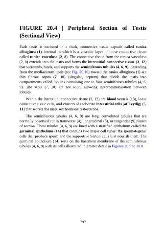
FIGURE  20.4  |  Peripheral  Section  of  Testis

               (Sectional View)


               Each  testis  is  enclosed  in  a  thick,  connective  tissue  capsule  called  tunica
               albuginea (1), internal to which is a vascular layer of loose connective tissue

               called tunica vasculosa (2, 8). The connective tissue from the tunica vasculosa
               (2, 8) extends into the testis and forms the interstitial connective tissue (3, 12)
               that surrounds, binds, and supports the seminiferous tubules (4, 6, 9). Extending

               from the mediastinum testis (see Fig. 20.10) toward the tunica albuginea (1) are
               thin  fibrous  septa  (7,  10)  (singular,  septum)  that  divide  the  testis  into
               compartments called lobules containing one to four seminiferous tubules (4, 6,
               9).  The  septa  (7,  10)  are  not  solid,  allowing  intercommunication  between
               lobules.


                   Within the interstitial connective tissue (3, 12) are blood vessels (13), loose
               connective tissue cells, and clusters of endocrine interstitial cells (of Leydig) (5,
               11) that secrete the male sex hormone testosterone.

                   The  seminiferous  tubules  (4,  6,  9)  are  long,  convoluted  tubules  that  are

               normally observed cut in transverse (4), longitudinal (6), or tangential (9) planes
               of section. These tubules (4, 6, 9) are lined with a stratified epithelium called the
               germinal epithelium (14) that contains two major cell types: the spermatogenic
               cells that produce sperm and the supportive Sertoli cells that nourish them. The
               germinal epithelium (14) rests on the basement membrane of the seminiferous

               tubules (4, 6, 9) with its cells illustrated in greater detail in Figures 20.5 to 20.8.


                                                          797