
Page 803 - Atlas of Histology with Functional Correlations
P. 803
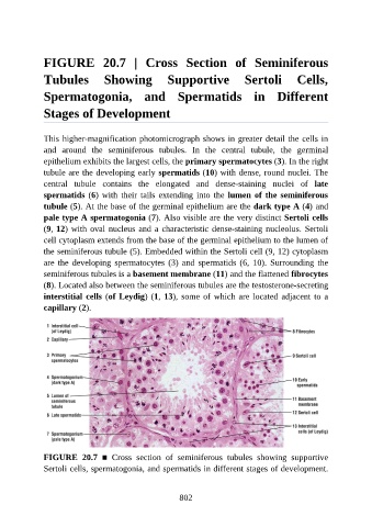
FIGURE  20.7  |  Cross  Section  of  Seminiferous

               Tubules  Showing  Supportive  Sertoli  Cells,

               Spermatogonia,  and  Spermatids  in  Different


               Stages of Development


               This higher-magnification photomicrograph shows in greater detail the cells in
               and  around  the  seminiferous  tubules.  In  the  central  tubule,  the  germinal

               epithelium exhibits the largest cells, the primary spermatocytes (3). In the right
               tubule are the developing early spermatids (10) with dense, round nuclei. The
               central  tubule  contains  the  elongated  and  dense-staining  nuclei  of  late
               spermatids (6) with their tails extending into the lumen of the seminiferous
               tubule (5). At the base of the germinal epithelium are the dark type A (4) and

               pale type A spermatogonia (7). Also visible are the very distinct Sertoli cells
               (9, 12) with oval nucleus and a characteristic dense-staining nucleolus. Sertoli
               cell cytoplasm extends from the base of the germinal epithelium to the lumen of

               the seminiferous tubule (5). Embedded within the Sertoli cell (9, 12) cytoplasm
               are  the  developing  spermatocytes  (3)  and  spermatids  (6,  10).  Surrounding  the
               seminiferous tubules is a basement membrane (11) and the flattened fibrocytes
               (8). Located also between the seminiferous tubules are the testosterone-secreting
               interstitial cells (of Leydig) (1, 13), some of which are located adjacent to a

               capillary (2).


               FIGURE  20.7  ■  Cross  section  of  seminiferous  tubules  showing  supportive
               Sertoli cells, spermatogonia, and spermatids in different stages of development.


                                                          802