
Page 806 - Atlas of Histology with Functional Correlations
P. 806
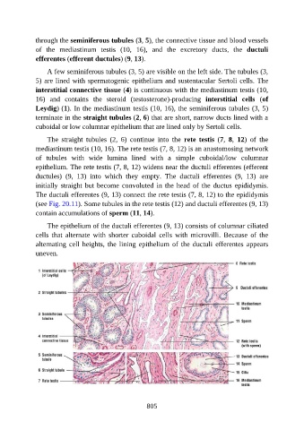
through the seminiferous tubules (3, 5), the connective tissue and blood vessels

               of  the  mediastinum  testis  (10,  16),  and  the  excretory  ducts,  the  ductuli
               efferentes (efferent ductules) (9, 13).

                   A few seminiferous tubules (3, 5) are visible on the left side. The tubules (3,
               5) are lined with spermatogenic epithelium and sustentacular Sertoli cells. The
               interstitial connective tissue (4) is continuous with the mediastinum testis (10,

               16)  and  contains  the  steroid  (testosterone)-producing  interstitial  cells  (of
               Leydig) (1). In the mediastinum testis (10, 16), the seminiferous tubules (3, 5)
               terminate in the straight tubules (2, 6) that are short, narrow ducts lined with a
               cuboidal or low columnar epithelium that are lined only by Sertoli cells.


                   The  straight  tubules  (2,  6)  continue  into  the  rete  testis  (7,  8,  12)  of  the
               mediastinum testis (10, 16). The rete testis (7, 8, 12) is an anastomosing network
               of  tubules  with  wide  lumina  lined  with  a  simple  cuboidal/low  columnar
               epithelium. The rete testis (7, 8, 12) widens near the ductuli efferentes (efferent
               ductules)  (9,  13)  into  which  they  empty.  The  ductuli  efferentes  (9,  13)  are

               initially straight but become convoluted in the head of the ductus epididymis.
               The ductuli efferentes (9, 13) connect the rete testis (7, 8, 12) to the epididymis
               (see Fig. 20.11). Some tubules in the rete testis (12) and ductuli efferentes (9, 13)

               contain accumulations of sperm (11, 14).

                   The epithelium of the ductuli efferentes (9, 13) consists of columnar ciliated
               cells  that  alternate  with  shorter  cuboidal  cells  with  microvilli.  Because  of  the
               alternating  cell  heights,  the  lining  epithelium  of  the  ductuli  efferentes  appears
               uneven.


                                                          805