
Page 810 - Atlas of Histology with Functional Correlations
P. 810
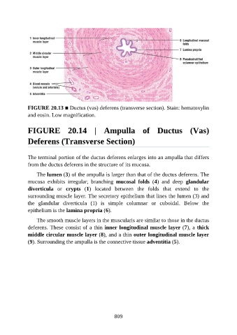
FIGURE 20.13 ■ Ductus (vas) deferens (transverse section). Stain: hematoxylin

               and eosin. Low magnification.


               FIGURE  20.14  |  Ampulla  of  Ductus  (Vas)


               Deferens (Transverse Section)


               The terminal portion of the ductus deferens enlarges into an ampulla that differs
               from the ductus deferens in the structure of its mucosa.

                   The lumen (3) of the ampulla is larger than that of the ductus deferens. The
               mucosa  exhibits  irregular,  branching  mucosal  folds  (4)  and  deep  glandular

               diverticula  or  crypts  (1)  located  between  the  folds  that  extend  to  the
               surrounding muscle layer. The secretory epithelium that lines the lumen (3) and
               the  glandular  diverticula  (1)  is  simple  columnar  or  cuboidal.  Below  the
               epithelium is the lamina propria (6).

                   The smooth muscle layers in the muscularis are similar to those in the ductus

               deferens. These consist of a thin inner longitudinal muscle layer (7), a thick
               middle circular muscle layer (8), and a thin outer longitudinal muscle layer
               (9). Surrounding the ampulla is the connective tissue adventitia (5).


                                                          809