
Page 809 - Atlas of Histology with Functional Correlations
P. 809
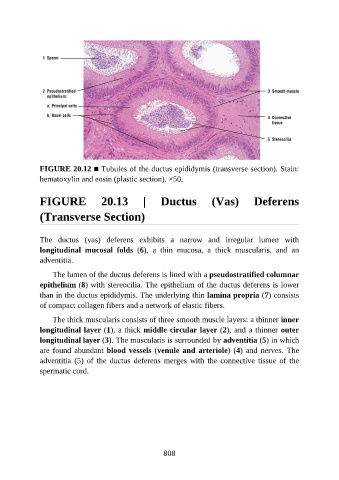
FIGURE 20.12 ■ Tubules of the ductus epididymis (transverse section). Stain:

               hematoxylin and eosin (plastic section). ×50.


               FIGURE                20.13         |     Ductus            (Vas)          Deferens


               (Transverse Section)


               The  ductus  (vas)  deferens  exhibits  a  narrow  and  irregular  lumen  with
               longitudinal  mucosal  folds  (6),  a  thin  mucosa,  a  thick  muscularis,  and  an
               adventitia.


                   The lumen of the ductus deferens is lined with a pseudostratified columnar
               epithelium (8) with stereocilia. The epithelium of the ductus deferens is lower
               than in the ductus epididymis. The underlying thin lamina propria (7) consists
               of compact collagen fibers and a network of elastic fibers.

                   The thick muscularis consists of three smooth muscle layers: a thinner inner

               longitudinal layer (1), a thick middle circular layer (2), and a thinner outer
               longitudinal layer (3). The muscularis is surrounded by adventitia (5) in which
               are found abundant blood vessels (venule and arteriole) (4)  and  nerves.  The
               adventitia  (5)  of  the  ductus  deferens  merges  with  the  connective  tissue  of  the

               spermatic cord.


                                                          808