
Page 804 - Atlas of Histology with Functional Correlations
P. 804
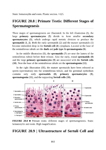
Stain: hematoxylin and eosin. Plastic section. ×125.


               FIGURE 20.8 | Primate Testis: Different Stages of

               Spermatogenesis


               Three  stages  of  spermatogenesis  are  illustrated.  In  the  left  illustration  (I),  the

               large  primary  spermatocytes  (5)  divide  to  form  smaller  secondary
               spermatocytes  (3),  which  undergo  rapid  meiotic  division  to  produce  the
               spermatids (1, 2). Both the early spermatids (2) and the mature spermatids (1)

               become embedded deep in the Sertoli cell (4) cytoplasm. Located at the base of
               the seminiferous tubule are the dark and pale type A spermatogonia (6).

                   In the middle illustration (II), the spermatids (7) are near the lumen of the
               seminiferous tubule before their release. Also the early, round spermatids (8)
               and the large primary spermatocytes (9) are associated with the Sertoli cells

               (10). Near the base of the seminiferous tubule are the spermatogonia (11).

                   In  the  right  illustration  (III),  the  mature  spermatids  have  been  released  as
               sperm (spermiation) into the seminiferous tubule, and the germinal epithelium
               contains  only  early  spermatids  (8),  primary  spermatocytes  (9),

               spermatogonia (11), and the supporting Sertoli cells (10).


               FIGURE  20.8  ■  Primate  testis:  different  stages  of  spermatogenesis.  Stain:
               hematoxylin and eosin. High magnification.


               FIGURE 20.9 | Ultrastructure of Sertoli Cell and


                                                          803