
Page 808 - Atlas of Histology with Functional Correlations
P. 808
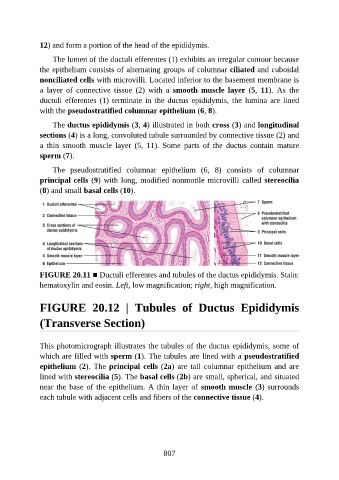
12) and form a portion of the head of the epididymis.

                   The lumen of the ductuli efferentes (1) exhibits an irregular contour because

               the epithelium consists of alternating groups of columnar ciliated and cuboidal
               nonciliated cells with microvilli. Located inferior to the basement membrane is
               a  layer  of  connective  tissue  (2)  with  a  smooth  muscle  layer  (5,  11).  As  the
               ductuli efferentes (1) terminate in the ductus epididymis, the lumina are lined

               with the pseudostratified columnar epithelium (6, 8).

                   The ductus epididymis (3, 4) illustrated in both cross (3) and longitudinal
               sections (4) is a long, convoluted tubule surrounded by connective tissue (2) and
               a  thin  smooth  muscle  layer  (5,  11).  Some  parts  of  the  ductus  contain  mature

               sperm (7).

                   The  pseudostratified  columnar  epithelium  (6,  8)  consists  of  columnar
               principal cells (9) with long, modified nonmotile microvilli called stereocilia
               (8) and small basal cells (10).


               FIGURE 20.11 ■ Ductuli efferentes and tubules of the ductus epididymis. Stain:
               hematoxylin and eosin. Left, low magnification; right, high magnification.


               FIGURE  20.12  |  Tubules  of  Ductus  Epididymis


               (Transverse Section)


               This photomicrograph illustrates the tubules of the ductus epididymis, some of
               which are filled with sperm (1). The tubules are lined with a pseudostratified

               epithelium (2). The principal cells (2a)  are  tall  columnar  epithelium  and  are
               lined with stereocilia (5). The basal cells (2b) are small, spherical, and situated
               near the base of the epithelium. A thin layer of smooth muscle (3) surrounds
               each tubule with adjacent cells and fibers of the connective tissue (4).


                                                          807