
Page 805 - Atlas of Histology with Functional Correlations
P. 805
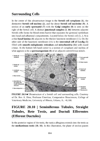
Surrounding Cells


               In the center of this ultrastructure image is the Sertoli cell cytoplasm (1), the
               distinctive Sertoli cell nucleus (2), and the dense Sertoli cell nucleolus (9). A
               section of an early spermatid (7) with the Golgi complex (8)  is  seen  on  the

               right of the Sertoli cell. A distinct junctional complex (3, 10) between adjacent
               Sertoli cells forms the blood–testis barrier that separates the germinal epithelium
               into basal and adluminal compartments. Located below the Sertoli cell (1, 2, 9) is
               a thin basal lamina (4) adjacent to the thicker basement membrane (11). On the
               other side of the basement membrane (11) is the interstitial cell of Leydig (5)

               filled with smooth endoplasmic reticulum and mitochondria (12) with round
               cristae. At the bottom left-hand corner is a section of cytoplasm and nucleus of
               what appears to be a spermatogonium (6) of an adjacent seminiferous tubule.


               FIGURE 20.9 ■ Ultrastructure of a Sertoli cell and surrounding cells. Courtesy
               of Dr. Rex A. Hess, Professor Emeritus, Comparative Biosciences, College of
               Veterinary Medicine, University of Illinois, Urbana, IL. ×8,100.


               FIGURE  20.10  |  Seminiferous  Tubules,  Straight

               Tubules,  Rete  Testis,  and  Ductuli  Efferentes


               (Efferent Ductules)


               In the posterior region of the testis, the tunica albuginea extends into the testis as
               the mediastinum testis (10, 16). In this illustration, the plane of section passes


                                                          804