
Page 800 - Atlas of Histology with Functional Correlations
P. 800
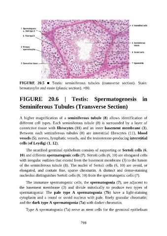
FIGURE  20.5  ■  Testis:  seminiferous  tubules  (transverse  section).  Stain:

               hematoxylin and eosin (plastic section). ×80.


               FIGURE  20.6  |  Testis:  Spermatogenesis  in


               Seminiferous Tubules (Transverse Section)


               A  higher  magnification  of  a  seminiferous  tubule  (8)  allows  identification  of
               different  cell  types.  Each  seminiferous  tubule  (8)  is  surrounded  by  a  layer  of
               connective tissue with fibrocytes (11) and an inner basement membrane (3).

               Between  each  seminiferous  tubules  (8)  are  interstitial  fibrocytes  (11),  blood
               vessels (5), nerves, lymphatic vessels, and the testosterone-producing interstitial
               cells (of Leydig) (1, 12).

                   The stratified germinal epithelium consists of supporting or Sertoli cells (6,
               10) and different spermatogenic cells (7). Sertoli cells (6, 10) are elongated cells

               with irregular outlines that extend from the basement membrane (3) to the lumen
               of the seminiferous tubule (8). The nuclei of Sertoli cells (6, 10) are ovoid, or
               elongated,  and  contain  fine,  sparse  chromatin.  A  distinct  and  dense-staining

               nucleolus distinguishes Sertoli cells (6, 10) from the spermatogenic cells (7).

                   The immature spermatogenic cells, the spermatogonia (7), are adjacent to
               the  basement  membrane  (3)  and  divide  mitotically  to  produce  two  types  of
               spermatogonia:  The  pale  type  A  spermatogonia  (7b)  have  a  light-staining
               cytoplasm and a round or ovoid nucleus with pale, finely granular chromatin;

               and the dark type A spermatogonia (7a) with darker chromatin.

                   Type A spermatogonia (7a) serve as stem cells for the germinal epithelium


                                                          799