
Page 799 - Atlas of Histology with Functional Correlations
P. 799
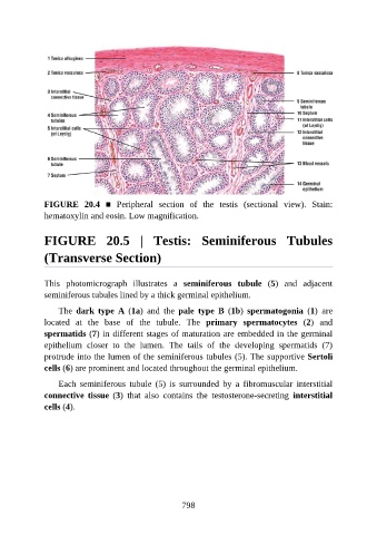
FIGURE  20.4  ■  Peripheral  section  of  the  testis  (sectional  view).  Stain:
               hematoxylin and eosin. Low magnification.


               FIGURE  20.5  |  Testis:  Seminiferous  Tubules


               (Transverse Section)


               This  photomicrograph  illustrates  a  seminiferous  tubule  (5)  and  adjacent
               seminiferous tubules lined by a thick germinal epithelium.

                   The  dark  type  A  (1a)  and  the  pale  type  B  (1b)  spermatogonia  (1)  are

               located  at  the  base  of  the  tubule.  The  primary  spermatocytes  (2)  and
               spermatids (7) in different stages of maturation are embedded in the germinal
               epithelium  closer  to  the  lumen.  The  tails  of  the  developing  spermatids  (7)

               protrude into the lumen of the seminiferous tubules (5). The supportive Sertoli
               cells (6) are prominent and located throughout the germinal epithelium.

                   Each  seminiferous  tubule  (5)  is  surrounded  by  a  fibromuscular  interstitial
               connective  tissue  (3)  that  also  contains  the  testosterone-secreting  interstitial
               cells (4).


                                                          798