
Page 88 - Atlas of Histology with Functional Correlations
P. 88
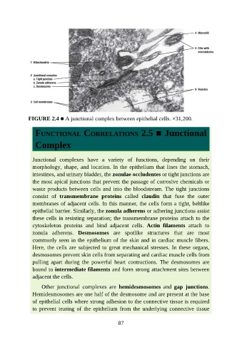
FIGURE 2.4 ■ A junctional complex between epithelial cells. ×31,200.


                  FUNCTIONAL CORRELATIONS 2.5 ■ Junctional


                  Complex


                 Junctional  complexes  have  a  variety  of  functions,  depending  on  their

                 morphology, shape,  and  location.  In  the epithelium  that  lines  the  stomach,
                 intestines, and urinary bladder, the zonulae occludentes or tight junctions are
                 the most apical junctions that prevent the passage of corrosive chemicals or

                 waste products between cells and into the bloodstream. The tight junctions
                 consist  of  transmembrane  proteins  called  claudin  that  fuse  the  outer
                 membranes of adjacent cells. In this manner, the cells form a tight, beltlike
                 epithelial barrier. Similarly, the zonula adherens or adhering junctions assist
                 these cells in resisting separation; the transmembrane proteins attach to the

                 cytoskeleton  proteins  and  bind  adjacent  cells.  Actin  filaments  attach  to
                 zonula  adherens.  Desmosomes  are  spotlike  structures  that  are  most
                 commonly seen in the epithelium of the skin and in cardiac muscle fibers.

                 Here,  the  cells  are  subjected  to  great  mechanical  stresses.  In  these  organs,
                 desmosomes prevent skin cells from separating and cardiac muscle cells from
                 pulling  apart  during  the  powerful  heart  contractions.  The  desmosomes  are
                 bound to intermediate filaments and form strong attachment sites between
                 adjacent the cells.


                     Other  junctional  complexes  are  hemidesmosomes  and  gap  junctions.
                 Hemidesmosomes are one half of the desmosome and are present at the base
                 of epithelial cells where strong adhesion to the connective tissue is required
                 to  prevent  tearing  of  the  epithelium  from  the  underlying  connective  tissue


                                                           87