
Page 92 - Atlas of Histology with Functional Correlations
P. 92
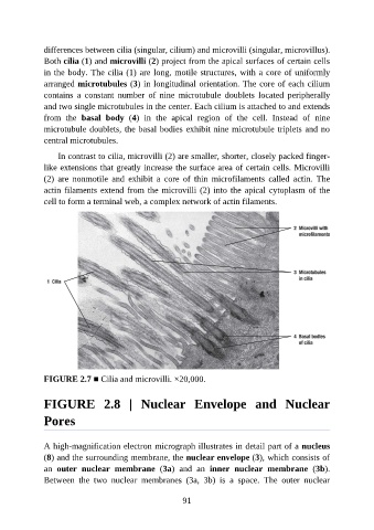
differences between cilia (singular, cilium) and microvilli (singular, microvillus).

               Both cilia (1) and microvilli (2) project from the apical surfaces of certain cells
               in the body. The cilia (1) are long, motile structures, with a core of uniformly
               arranged microtubules (3) in longitudinal orientation. The core of each cilium
               contains  a  constant  number  of  nine  microtubule  doublets  located  peripherally
               and two single microtubules in the center. Each cilium is attached to and extends

               from  the  basal  body  (4)  in  the  apical  region  of  the  cell.  Instead  of  nine
               microtubule doublets, the basal bodies exhibit nine microtubule triplets and no
               central microtubules.

                   In contrast to cilia, microvilli (2) are smaller, shorter, closely packed finger-

               like extensions that greatly increase the surface area of certain cells. Microvilli
               (2)  are  nonmotile  and  exhibit  a  core  of  thin  microfilaments  called  actin.  The
               actin filaments extend from the microvilli (2) into the apical cytoplasm of the
               cell to form a terminal web, a complex network of actin filaments.


               FIGURE 2.7 ■ Cilia and microvilli. ×20,000.


               FIGURE  2.8  |  Nuclear  Envelope  and  Nuclear


               Pores


               A high-magnification electron micrograph illustrates in detail part of a nucleus
               (8) and the surrounding membrane, the nuclear envelope (3), which consists of
               an  outer  nuclear  membrane  (3a)  and  an  inner  nuclear  membrane  (3b).

               Between  the  two  nuclear  membranes  (3a,  3b)  is  a  space.  The  outer  nuclear


                                                           91