
Page 96 - Atlas of Histology with Functional Correlations
P. 96
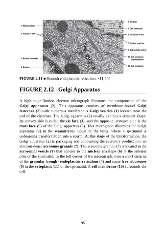
FIGURE 2.11 ■ Smooth endoplasmic reticulum. ×11,500.


               FIGURE 2.12 | Golgi Apparatus


               A  high-magnification  electron  micrograph  illustrates  the  components  of  the
               Golgi  apparatus  (2).  This  apparatus  consists  of  membrane-bound  Golgi

               cisternae (2)  with  numerous  membranous  Golgi vesicles (1)  located  near  the
               end of the cisternae. The Golgi apparatus (2) usually exhibits a crescent shape.
               Its convex side is called the cis face (3), and the opposite, concave side is the

               trans face (9) of the Golgi apparatus (2). This micrograph illustrates the Golgi
               apparatus  (2)  in  the  seminiferous  tubule  of  the  testis,  where  a  spermatid  is
               undergoing transformation into a sperm. At this stage of the transformation, the
               Golgi apparatus (2) is packaging and condensing the secretory product into an
               electron-dense acrosome granule (7). The acrosome granule (7) is located in the

               acrosomal vesicle (8) that adheres to the nuclear envelope (6) at the anterior
               pole of the spermatid. In the left corner of the micrograph, note a short cisterna
               of the granular (rough) endoplasmic reticulum (4) and some free ribosomes

               (5) in the cytoplasm (11) of the spermatid. A cell membrane (10) surrounds the
               cell.


                                                           95