
Page 97 - Atlas of Histology with Functional Correlations
P. 97
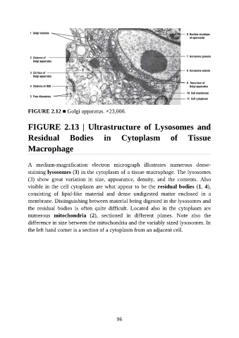
FIGURE 2.12 ■ Golgi apparatus. ×23,000.


               FIGURE  2.13  |  Ultrastructure  of  Lysosomes  and

               Residual  Bodies  in  Cytoplasm  of  Tissue

               Macrophage


               A  medium-magnification  electron  micrograph  illustrates  numerous  dense-
               staining lysosomes (3) in the cytoplasm of a tissue macrophage. The lysosomes
               (3)  show  great  variation  in  size,  appearance,  density,  and  the  contents.  Also
               visible in the cell cytoplasm are what appear to be the residual bodies (1, 4),

               consisting  of  lipid-like  material  and  dense  undigested  matter  enclosed  in  a
               membrane. Distinguishing between material being digested in the lysosomes and
               the  residual  bodies  is  often  quite  difficult.  Located  also  in  the  cytoplasm  are
               numerous  mitochondria  (2),  sectioned  in  different  planes.  Note  also  the

               difference in size between the mitochondria and the variably sized lysosomes. In
               the left hand corner is a section of a cytoplasm from an adjacent cell.


                                                           96