
Page 94 - Atlas of Histology with Functional Correlations
P. 94
Nuclear pores control the transport of macromolecules between the
nucleus and the cytoplasm. The nuclear pore membrane, like other cell
membranes, shows selective permeability. As a result, some of the larger
molecules travel through the pores via an active transport mechanism.
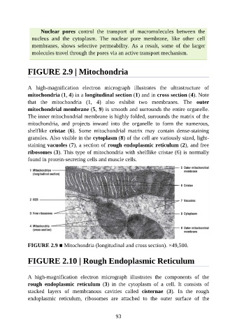
FIGURE 2.9 | Mitochondria
A high-magnification electron micrograph illustrates the ultrastructure of
mitochondria (1, 4) in a longitudinal section (1) and in cross section (4). Note
that the mitochondria (1, 4) also exhibit two membranes. The outer
mitochondrial membrane (5, 9) is smooth and surrounds the entire organelle.
The inner mitochondrial membrane is highly folded, surrounds the matrix of the
mitochondria, and projects inward into the organelle to form the numerous,
shelflike cristae (6). Some mitochondrial matrix may contain dense-staining
granules. Also visible in the cytoplasm (8) of the cell are variously sized, light-
staining vacuoles (7), a section of rough endoplasmic reticulum (2), and free
ribosomes (3). This type of mitochondria with shelflike cristae (6) is normally
found in protein-secreting cells and muscle cells.
FIGURE 2.9 ■ Mitochondria (longitudinal and cross section). ×49,500.
FIGURE 2.10 | Rough Endoplasmic Reticulum
A high-magnification electron micrograph illustrates the components of the
rough endoplasmic reticulum (3) in the cytoplasm of a cell. It consists of
stacked layers of membranous cavities called cisternae (3). In the rough
endoplasmic reticulum, ribosomes are attached to the outer surface of the
93