
Page 90 - Atlas of Histology with Functional Correlations
P. 90
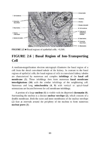
FIGURE 2.5 ■ Basal regions of epithelial cells. ×9,500.


               FIGURE  2.6  |  Basal  Region  of  Ion-Transporting

               Cell


               A  medium-magnification  electron  micrograph  illustrates  the  basal  region  of  a
               cell  from  the  distal  convoluted  tubule  of  the  kidney.  In  contrast  to  the  basal
               regions of epithelial cells, the basal regions of cells in convoluted kidney tubules
               are  characterized  by  numerous  and  complex  infoldings  of  the  basal  cell

               membrane  (5).  These  infoldings  then  form  numerous  basal  membrane
               interdigitations  (10)  with  the  similar  infoldings  of  the  neighboring  cell.
               Numerous  and  long  mitochondria  (4,  9)  with  vertical  or  apical–basal
               orientations are located between the cell membrane infoldings.

                   A portion of a large nucleus (1) is visible with its dispersed chromatin (8).

               Surrounding the nucleus is a distinct nuclear envelope (2), which consists of a
               double membrane. Both the outer and inner membranes of the nuclear envelope
               (2)  fuse  at  intervals  around  the  periphery  of  the  nucleus  to  form  numerous

               nuclear pores (3).


                                                           89