
Page 116 - C:\Users\uromn\Videos\seyyedi pdf\
P. 116
Dalirsani and Seyyedi: CO2 laser on plaque‑like OLP
Subjects was seen on the right buccal mucosa. Two months later,
Patients with plaque-like OLP, being confirmed by mild keratotic striae were observed [Figure 1c]. In that
clinical and histhopathological evaluations, were enrolled session, another plaque-like lesion of size 20 × 8 mm on
to this study. The patients were excluded from the study the left buccal mucosa was under laser irritation with
if any dysplasia was observed in the tissue samples, or the same properties.
they were receiving immunosuppressive drugs, or if they The patient referred about 10 years later. He had not
had any systemic diseases, or were pregnant. used any drug during these years and did not suffer
Laser properties from burning on the laser therapy region.
After local anesthesia, all plaque-like lesions were The plaque form of OLP was not observed on the right
evaporated with a CO2 laser system (Ultra dream buccal mucosa; however, there was a mild atrophic patch
pulsed laser DS-40UB; Korea). The power of 4–7 watt about 20 × 10 mm associated with reticular white striae
as continuous wave (CW) and defocused mode with spot without any burning [Figure 1d]. On the left buccal
size of 0.2 mm was used to evaporate the entire plaque- mucosa, a thin white plaque with size of 10 × 5 mm was
like lesions. After operation, NSAIDs were prescribed for seen [Figure 1e].
relieving the pain. On the right border of tongue, a mild keratotic lesion and
Ethical approval atrophic patch were observed, which were associated with
All patients signed written informed consent forms. mild burning. He was advised to use dexamethasone and
The protocol of this study was approved in 2018 by nystatin mouthwashes for atrophic lesions. After 3 weeks,
ethical committee of Mashhad University of Medical a noticeable improvement in theses lesions occurred.
Sciences (the ethical code: IR.MUMS.DENTISTRY. Patient no. 2
REC.900098). A 40-year-old female referred with a complaint of
Patient No. 1 burning since 2 years ago. In the clinical examination,
there was a keratotic plaque with size of 25 × 20 mm on
A 45-year-old man was referred to the dental school the left buccal mucosa. Also, keratotic reticular lesion
with a keratotic plaque lesion of size 49 × 11 mm on was observed on the left lateral border of the tongue.
the right buccal mucosa. The patient mentioned that he
saw this white lesion about one year ago, although he The buccal lesion was evaporated with continuous mode
did not complain of any pain or burning. An incisional of CO2 laser using a power of 7 watt. The irradiation
biopsy performed and histopathological evaluation continued until removing the plaque-like lesion.
showed OLP. Follow‑up sessions
The lesion was evaporated with CW and defocused CO2 The map-like ulcer coated with pseudomembrane was
laser with a power of 7 watt. The irradiation continued observed one week later. The patient reported mild
until removing the white plaque [Figure 1a]. burning. Three months later, mild keratotic reticular
striae were seen on the left buccal mucosa. The patient
Follow‑up sessions was prescribed topical dexamethasone and nystatin
In follow-up visit at one week later, the map-like ulcer mouthwashes.
coated with pseudomembrane was observed in the
operation site [Figure 1b]. The patient reported mild Four months later, mild keratotic and atrophic lesions
burning. About 40 days later, a mild keratotic lesion were observed on the buccal mucosa and lateral border
of the tongue. Topical treatment was continued.
During one year later, with regular visits every 2 months,
no recurrence of plaque lesion was seen; however, there
were mild keratotic and atrophic lesions on the tongue
and buccal mucosa.
a b Patient no. 3
A 34-year-old man referred to the dental school
complaining of white patch on his buccal mucosa since
2 years ago.
c d e In clinical examination, a keratotic plaque-like lesion
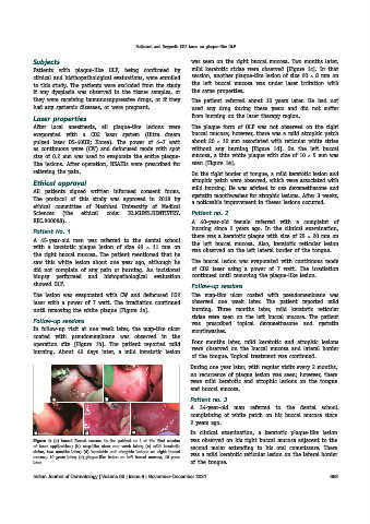
Figure 1: (a) buccal Buccal mucosa in the patient no.1 at the first session was observed on his right buccal mucosa adjacent to the
of laser application; (b) map-like ulcer one week later; (c) mild keratotic second molar extending to his oral commissure. There
striae, two months later; (d) keratotic and atrophic lesions on right buccal was a mild keratotic reticular lesion on the lateral border
mucosa, 10 years later; (e) plaque-like lesion on left buccal mucosa, 10 years
later of the tongue.
Indian Journal of Dermatology | Volume 66 | Issue 6 | November-December 2021 699