
Page 117 - C:\Users\uromn\Videos\seyyedi pdf\
P. 117
Dalirsani and Seyyedi: CO2 laser on plaque‑like OLP
Continuous mode of CO2 laser with a power of 5 of OLP through incisional biopsy and pathological
watt was used for excision of the plaque in two assessment, the plaque-like lesion was irradiated with
sessions [Figure 2]. CO2 laser [Figure 3b].
Follow‑up sessions Follow‑up sessions
After one week, a map-like ulcer with erythematous halo Two weeks later, the ulcer healed partially. The patient
was seen in the operation site. The patient suffered from reported mild burning and was satisfied with the
moderate burning. Diphenhydramine mouthwash was improvement process.
prescribed for pain relief.
The patient was under examination every 6 months for
Two weeks later, there were ulcer scar and mild keratotic 4 years. In this part of buccal mucosa, no plaque-like
striae at the site of laser operation; the patient did not lesion was observed.
report any burning sensation. Patient no. 6
Patient no. 4 A 62-year-old woman referred to a dental clinic with a
A 77- year-old woman referred to the dental school complaint of white lesions on alveolar mucosa and floor
complaining of burning tongue since 4 years ago. of mouth and lateral border of her tongue [Figure 4a].
After incisional biopsy and histopathological
There was a 30 × 10 mm keratotic plaque on the assessment, OLP was diagnosed. At the first session,
right border of her tongue. Also, there were bilaterally CO2 laser was employed for surgery of plaque-like
atrophic and keratotic lesions with Wickham’s striae on lesions of alveolar ridge and some parts of floor of
her tongue and buccal mucosa. mouth [Figure 4b]. After 2 weeks, the rest of lesions
For excision of the keratotic plaque, defocused continuous on floor of mouth and ventral surface of her tongue
CO2 laser with a power of 4–7 watt was employed. The were removed with laser.
irradiation continued to evaporate the plaque-like lesion Follow‑up sessions
completely.
The follow-up of the patient was continued every
Follow‑up sessions 2 months. No recurrence occurred until 8 months later.
After 20 days, the irregular wound of size 20 × 10 mm Patient no. 7
was observed. Because of some atrophic lesions on her A 60-year-old man with plaque-like and reticular white
tongue and buccal mucosa, the patient was prescribed lesions on his left maxillary ridge and hard palatal
topical dexamethasone and nystatin mouthwashes. mucosa referred to a dental clinic. He did not suffer
Patient no. 5 from any burning. CO2 laser was used for evaporation of
A 30 × 10 mm keratotic plaque was observed on the plaque-like lesion.
right buccal mucosa extended to commissure region Follow‑up sessions
in a 36-year-old man [Figure 3a]. There were mild After 2 weeks, the ulcer due to laser irradiation on the
keratotic striae around the plaque. The patient had ridge was partially epithelized. During 2-year follow-up
seen the lesions 14 months ago and complained of examinations, recurrence did not happen.
moderate burning. After confirming the diagnosis
Patient no. 8
A keratotic plaque was observed on floor of mouth in
a 30-year-old man. The clinical examination revealed
an extensive keratotic plaque with different thickness.
Bilaterally keratotic lesions were seen on ventral surface
of his tongue and the right lingual alveolar mucosa and
a b
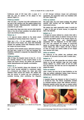
Figure 2: Buccal mucosa in the patient no. 3; one week after laser therapy; map- Figure 3: (a) Keratotic plaque on right buccal mucosa in the patient no. 5. (b)
like ulcer was seen carbonization layer immediately after laser evaporation
700 Indian Journal of Dermatology | Volume 66 | Issue 6 | November-December 2021