
Page 36 - DP Vol 20 No 5_Neat
P. 36
COSMETIC SECTION
Fig 10
Fig 10-11: Laboratory work
Fig 11
Fig 12.1: Mandibular preparations
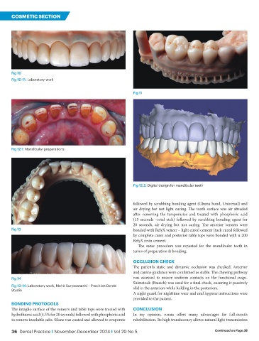
Fig 12.2: Digital design for mandibular teeth
followed by scrubbing bonding agent (Gluma bond, Universal) and
air drying but not light curing. The tooth surface was air abraded
after removing the temporaries and treated with phosphoric acid
(15 seconds –total etch) followed by scrubbing bonding agent for
20 seconds, air drying but not curing. The anterior veneers were
Fig 13 bonded with RelyX veneer – light cured cement (tack cured followed
by complete cure) and posterior table tops were bonded with u 200
RelyX resin cement.
The same procedure was repeated for the mandibular teeth in
terms of preparation & bonding.
OCCLUSION CHECK
The patient’s static and dynamic occlusion was checked. Anterior
and canine guidance were confirmed as stable. The chewing pathway
Fig 14 was assessed to ensure uniform contacts on the functional cusps.
Shimstock (Bausch) was used for a final check, ensuring it passively
Fig 13-14: Laboratory work, Mohit Suryavanashi – Precision Dental slid in the anteriors while holding in the posteriors.
Studio
A night guard for nighttime wear and oral hygiene instructions were
provided to the patient.
BONDING PROTOCOLS
The intaglio surface of the veneers and table tops were treated with CONCLUSION
hydrofluoric acid (4.5% for 20 seconds) followed with phosphoric acid In my opinion, e.max offers many advantages for full-mouth
to remove insoluble salts. Silane was coated and allowed to evaporate rehabilitation. Its high translucency allows natural light transmission
36 Dental Practice I November-December 2024 I Vol 20 No 5 Continued on Page 38