
Page 101 - Power of Stem Cells- arthritis and regeneration
P. 101
Liu et al. Arthritis Research & Therapy 2010, 12:R210 Page 10 of 13
http://arthritis-research.com/content/12/6/R210
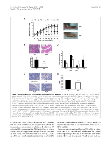
Figure 6 UC-MSCs prevented tissue damage and inflammatory responses in CIA. (a) Treatment was begun after the onset of disease
6
(arthritis score≥1). PBS and PBS containing 1 × 10 UC-MSCs were injected intraperitoneally each day for five days to mice with CIA. The severity
of CIA was progressively attenuated in UC-MSCs treated mice, as compared with PBS treated mice. N = 10, **P < 0.01, vs. the PBS controls. All
the data are expressed as the mean ± SD. (b) H & E-stained sagittal sections of joints from CIA mice. PBS treated mice showed a marked
mononuclear cell infiltration, severe synovitis, pannus formation and bone erosion. However, the majority of joints from mice injected with
UC-MSCs had normal morphology with a smooth articulation cartilage surface, and an absence of inflammatory cell infiltrate and pannus
formation. Original magnification × 100. N = 10, **P < 0.01, vs. the PBS controls. All the data are expressed as the mean ± SD. (c) UC-MSCs
treatment reduced inflammatory responses in CIA. There were reduced levels of proinflammatory cytokines and chemokines (TNF-a, IL-6 and
MCP-1) and increased levels of the anti-inflammatory/regulatory cytokine (IL-10) in sera of UC-MSC-treated mice, in comparison with PBS treated
mice. N = 10, ** P < 0.01, * P < 0.05, respectively. All the data are expressed as the mean ± SD. (d) UC-MSCs were detected in the spleen of CIA
mice. mAb against human nuclei was used to detect human UC-MSCs in CIA mice, on Day 3 and Day 7, UC-MSCs were detected in the spleen.
Arrows indicate human UC-MSCs in the spleen. Original magnification × 200. (e) DTH responses in UC-MSC-treated or untreated CII immunized
mice. Values are the mean ± SD. N =5.
CII-activated PBMCs from RA patients [17]. However, mediated T-cell inhibition, while IDO, TGF-b1 and IL-10
the TGFb1 blockade did not significantly affect the were mainly involved in the suppressive effect of UC-
immunosuppressive action of hASCs on T cells from RA MSCs on FLSs.
patients [18], suggesting that MSCs of different origins Systemic administration of human UC-MSCs in estab-
maybe mediated suppression through different cytokines. lished CIA in mice significantly ameliorated the clinical
In this study, we demonstrated that TGF-b1, PGE2 and histopathologic severity of the disease. The thera-
and NO are potent modulators involved in UC-MSCs peutic effect was xenogeneic, which means that the