
Page 96 - Power of Stem Cells- arthritis and regeneration
P. 96
Liu et al. Arthritis Research & Therapy 2010, 12:R210 Page 5 of 13
http://arthritis-research.com/content/12/6/R210
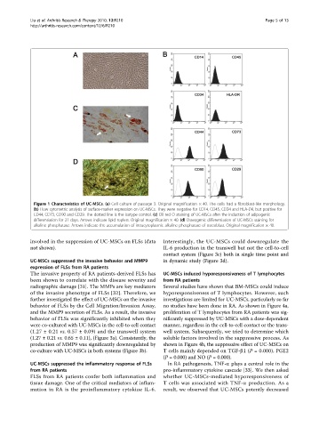
Figure 1 Characteristics of UC-MSCs. (a) Cell culture of passage 3. Original magnification × 40. The cells had a fibroblast-like morphology.
(b) Flow cytometric analysis of surface-marker expression on UC-MSCs. They were negative for CD14, CD45, CD34 and HLA-DR, but positive for
CD44, CD73, CD90 and CD29. The dotted line is the isotype control. (c) Oil red O staining of UC-MSCs after the induction of adipogenic
differentiation for 21 days. Arrows indicate lipid roplets. Original magnification × 40. (d) Osteogenic differentiation of UC-MSCs staining for
alkaline phosphatase. Arrows indicate the accumulation of intracytoplasmic alkaline phosphatase of osteoblast. Original magnification × 40.
involved in the suppression of UC-MSCs on FLSs (data Interestingly, the UC-MSCs could downregulate the
not shown). IL-6 production in the transwell but not the cell-to-cell
contact system (Figure 3c) both in single time point and
UC-MSCs suppressed the invasive behavior and MMP9 in dynamic study (Figure 3d).
expression of FLSs from RA patients
The invasive property of RA patients-derived FLSs has UC-MSCs induced hyporesponsiveness of T lymphocytes
been shown to correlate with the disease severity and from RA patients
radiographic damage [31]. The MMPs are key mediators Several studies have shown that BM-MSCs could induce
of the invasive phenotype of FLSs [32]. Therefore, we hyporesponsiveness of T lymphocytes. However, such
further investigated the effect of UC-MSCs on the invasive investigations are limited for UC-MSCs, particularly so far
behavior of FLSs by the Cell Migration/Invasion Assay, no studies have been done in RA. As shown in Figure 4a,
and the MMP9 secretion of FLSs. As a result, the invasive proliferation of T lymphocytes from RA patients was sig-
behavior of FLSs was significantly inhibited when they nificantly suppressed by UC-MSCs with a dose-dependent
were co-cultured with UC-MSCs in the cell-to-cell contact manner, regardless in the cell-to-cell contact or the trans-
(1.27 ± 0.21 vs. 0.57 ± 0.09) and the transwell system well system. Subsequently, we tried to determine which
(1.27 ± 0.21 vs. 0.65 ± 0.11), (Figure 3a). Consistently, the soluble factors involved in the suppressive process. As
production of MMP9 was significantly downregulated by shown in Figure 4b, the suppressive effect of UC-MSCs on
co-culture with UC-MSCs in both systems (Figure 3b). T cells mainly depended on TGF-b1(P =0.000), PGE2
(P = 0.000) and NO (P = 0.000).
UC-MSCs suppressed the inflammatory response of FLSs In RA pathogenesis, TNF-a plays a central role in the
from RA patients pro-inflammatory cytokine cascade [33]. We then asked
FLSs from RA patients confer both inflammation and whether UC-MSCs-mediated hyporesponsiveness of
tissue damage. One of the critical mediators of inflam- T cells was associated with TNF-a production. As a
mation in RA is the proinflammatory cytokine IL-6. result, we observed that UC-MSCs potently decreased