
Page 99 - Power of Stem Cells- arthritis and regeneration
P. 99
Liu et al. Arthritis Research & Therapy 2010, 12:R210 Page 8 of 13
http://arthritis-research.com/content/12/6/R210
the activation of autoreactive Th1 cells [37,38]. Downre-
gulation of the inflammatory Th1 and the elevated IL-10
levels by UC-MSCs prompted us to further investigate
the effect of Tregs in immunosuppressant action of
UC-MSCs in vivo.Asshown in Figure 7d, wefound that
+
there were significantly higher numbers of CD4 Foxp3 +
Tregs in spleen and peripheral blood in the UC-MSC-
treated mice than the PBS treated mice. Moreover,
+
+
CD4 CD25 T cells isolated from human UC-MSC-trea-
ted mice functioned as suppressive Treg cells, since they
inhibited the proliferation of syngeneic T cells stimulated
with CD3 and CD28 (Figure 7e).
Discussion
In the present study, we provided evidence that
UC-MSCs can exert a profound inhibitory effect on FLSs
and T cells from RA patients. They could suppress prolif-
eration, the invasive behavior and inflammatory
responses of FLSs, inhibit activation of T cells and induce
the Tregs expression. Furthermore, we showed that
UC-MSC mediated suppression on T cells and FLSs pro-
liferation through several soluble factors, including IDO,
PGE2, NO, IL-10 and TGF-b1, respectively. Systemic
infusion of UC-MSCs significantly reduced the severity
of CIA in mice. The improvement of clinical manifesta-
tion was accompanied by the decreased secretion of var-
ious inflammatory cytokines and chemokines, and the
downregulated Th1/Th17 cells. Furthermore, in the
UC-MSCs treated mice, the expansion of Th2/Tregs and
the production of anti-inflammatory IL-10 were elevated.
MSCs have the capability of self-renewal and differen-
tiation into various lineages of mesenchymal tissues.
Moreover, MSCs have been consistently shown to exert a
potent immunosuppressive effect superior in magnitude
to any other immunosuppressive cell types thus far
described [39]. Compared with those from bone marrow,
MSCs derived from UC have higher proliferative potency,
stronger differentiation capacity, and lower risk for viral
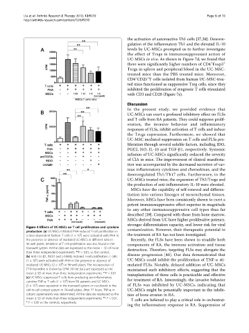
Figure 4 Effects of UC-MSCs on T cell proliferation and cytokine
production. (a) UC-MSCs inhibited PHA-induced T-cell proliferation in contamination. However, their therapeutic potential in
5
a dose-dependent fashion. T cells (1 × 10 ) were activated with PHA in the treatment of RA has not been investigated.
the presence or absence of irradiated UC-MSCs in different ratio in Recently, the FLSs have been shown to straddle both
96-well plates. Inhibition of T cell proliferation was also found in the components of RA, the immune activation and tissue
transwell system. All the data are expressed as the mean ± SD of more destruction. Therefore, targeting FLSs may abrogate the
than three independent experiments. **P < 0.01, vs. the control.
(b) Anti-TGF-b1, INDO and L-NAME restored T-cell proliferation. T cells disease progression [40]. Our data demonstrated that
5
(1 × 10 ) were activated with PHA in the presence or absence of UC-MSCs could inhibit the proliferation of TNF-a sti-
4
irradiated UC-MSCs (2 × 10 ) in 96-well plates. The incorporation of mulated FLSs. Notably, delayed addition of UC-MSCs
3
( H)-thymidine is shown by CPM. All the data are expressed as the maintained such inhibitory effects, suggesting that the
mean ± SD of more than three independent experiments. **P < 0.01. transplantation of these cells is practicable and effective
(c) UC-MSCs suppressed T cells from producing pro-inflammatory
6
cytokine TNF-a. T cells (1 × 10 ) from RA patients and UC-MSCs for treatment of RA. Interestingly, the invasive behavior
4
(5 × 10 ) were separated in the transwell system or cocultured in the of FLSs was inhibited by UC-MSCs, indicating that
cell-to-cell contact system in 24-well plates. After 72 hours, TNF-a in UC-MSCs might be potentially important in the inhibi-
culture supernatants was determined. All the data are expressed as the tion of bone erosion in RA.
mean ± SD of more than three independent experiments. ** P <0.01,
T cells are believed to play a critical role in orchestrat-
* P < 0.05 vs. the controls, respectively.
ing the inflammatory response in RA. Suppression of