
Page 97 - Power of Stem Cells- arthritis and regeneration
P. 97
Liu et al. Arthritis Research & Therapy 2010, 12:R210 Page 6 of 13
http://arthritis-research.com/content/12/6/R210
the production of TNF-a, both in the cell-to-cell con-
tact and the transwell system, especially in PHA acti-
vated T cells (Figure 4c).
UC-MSCs induced Tregs from RA patients
Given the concept that Tregs play a critical role in the
maintenance of self-immune tolerance in RA [34],
UC-MSCs exert an immunoregulatory function on FLSs
and T cells. The next intriguing question is whether
UC-MSCs play a role in the induction of Tregs in RA.
+
Recent studies demonstrated that not all CD4 CD25 bright
+
cells coexpressed Foxp3, while some Foxp3 cells resided
-
in the CD25 dim or CD25 population [35]. In this study,
+
+
+
the expression of FoxP3 on CD4 T cells (CD4 Foxp3 )
was defined as Tregs. Notably, the percentages of CD4 +
+
Foxp3 T cells were significantly higher in the presence
of UC-MSCs, irrespective of PHA stimulation (Figure 5).
UC-MSCs prevented tissue damage in CIA
The immunosuppressive effects of UC-MSCs on T cells
and FLSs in human RA promoted us to investigate the
potential therapeutic effects of UC-MSCs in CIA, which
is an arthritis model that shares a number of clinical,
histologic and immunologic features of RA. As shown in
Figure 6a, the severity of CIA was progressively attenu-
ated in UC-MSCs treated mice, as compared with PBS
treated mice. Moreover, the therapeutic effect was speci-
fic to viable human UC-MSCs, because dead human
UC-MSCs and human FLSs from traumatic patients
without arthritis failed to prevent the progression of
arthritis. The therapeutic effects of UC-MSCs on CIA in
mice were further verified by histological examination at
the endpoint of clinical study. We observed that control
mice exhibited a marked mononuclear cell infiltration,
severe synovitis, pannus formation and bone erosion. In
contrast, the majority of joints from mice injected with
UC-MSCs had normal morphology with a smooth
articulation cartilage surface, and an absence of inflam-
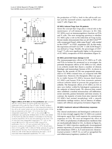
Figure 2 Effects of UC-MSCs on FLSs proliferation. (a) Compared
matory cell infiltrate and pannus formation (Figure 6b).
with the control, TNF-a (20 ng/ml) significantly induced the
proliferation of FLSs after five days of culture. UC-MSCs inhibited
TNF-a-stimulated-FLSs proliferation in a dose-dependent fashion in UC-MSCs treatment reduced inflammatory responses
the cell-to-cell contact system and also the transwell system. All the in CIA
data are expressed as the mean ± SD of more than three The clinical amelioration and histological verification in
independent experiments. **P < 0.01 vs. the controls. (b) FLSs
proliferation was significantly inhibited when UC-MSCs were added CIA in mice strongly suggests that UC-MSCs are a potent
on the fourth day after the initiation of stimulation in the five-day tolerogenic agent that could suppress the autoimmune
coculture experiment. All the data are expressed as the mean ± SD responses in CIA. We next investigated the effect of
of more than three independent experiments. **P < 0.01 vs. the UC-MSCs on production of inflammatory mediators that
control. (c) Anti-IL-10, 1-MT and anti-TGF-b1 restored FLSs are mechanistically linked to CIA. As shown in Figure 6c,
4
proliferation. FLSs (1 × 10 ) were activated with TNF-a in the
4
presence or absence of irradiated MSCs (1 × 10 ) in 96-well plates. human UC-MSCs injection significantly downregulated
Anti-IL-10 (10 μg/Ml), 1-MT (1 mM) and TGF-b1 antibody (10 μg/mL) protein expression of various proinflammatory cytokines
3
were added for five days. The incorporation of ( H)-thymidine is and chemokines (TNF-a, IL-6 and monocyte chemoat-
shown by CPM. All the data are expressed as the mean ± SD of tractant protein-1 (MCP-1)), as well as upregulted the
more than three independent experiments. **P < 0.01.
anti-inflammatory/regulatory cytokine (IL-10).