
Page 98 - Power of Stem Cells- arthritis and regeneration
P. 98
Liu et al. Arthritis Research & Therapy 2010, 12:R210 Page 7 of 13
http://arthritis-research.com/content/12/6/R210
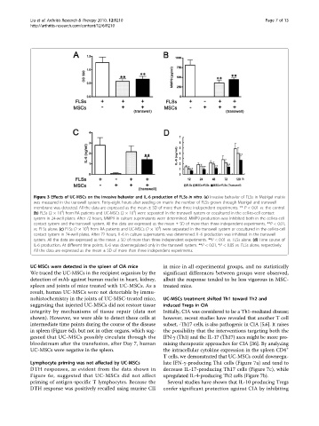
Figure 3 Effects of UC-MSCs on the invasive behavior and IL-6 production of FLSs in vitro. (a) Invasive behavior of FLSs in Matrigel matrix
was measured in the transwell system. Forty-eight hours after seeding on matrix the number of FLSs grown through Matrigel and transwell
membrane was detected. All the data are expressed as the mean ± SD of more than three independent experiments. ** P < 0.01 vs. the control.
4
4
(b) FLSs (2 × 10 ) from RA patients and UC-MSCs (2 × 10 ) were separated in the transwell system or cocultured in the cell-to-cell contact
system in 24-well plates. After 72 hours, MMP9 in culture supernatants were determined. MMP9 production was inhibited both in the cell-to-cell
contact system and the transwell system. All the data are expressed as the mean ± SD of more than three independent experiments. **P < 0.01,
4
4
vs. FLSs alone. (c) FLSs (2 × 10 ) from RA patients and UC-MSCs (2 × 10 ) were separated in the transwell system or cocultured in the cell-to-cell
contact system in 24-well plates. After 72 hours, IL-6 in culture supernatants was determined. IL-6 production was inhibited in the transwell
system. All the data are expressed as the mean ± SD of more than three independent experiments. **P < 0.01 vs. FLSs alone. (d) Time course of
IL-6 production. At different time points, IL-6 was downregulated only in the transwell system. **P < 0.01, *P < 0.05 vs. FLSs alone, respectively.
All the data are expressed as the mean ± SD of more than three independent experiments.
UC-MSCs were detected in the spleen of CIA mice in mice in all experimental groups, and no statistically
We traced the UC-MSCs in the recipient organism by the significant differences between groups were observed,
detection of mAb against human nuclei in heart, kidney, albeit the response tended to be less vigorous in MSC-
spleen and joints of mice treated with UC-MSCs. As a treated mice.
result, human UC-MSCs were not detectable by immu-
nohistochemistry in the joints of UC-MSC-treated mice, UC-MSCs treatment shifted Th1 toward Th2 and
suggesting that injected UC-MSCs did not restore tissue induced Tregs in CIA
integrity by mechanisms of tissue repair (data not Initially, CIA was considered to be a Th1-mediated disease;
shown).However,wewereabletodetect these cellsat however, recent studies have revealed that another T cell
intermediate time points during the course of the disease subset, -Th17 cells, is also pathogenic in CIA [5,6]. It raises
in spleen (Figure 6d), but not in other organs, which sug- the possibility that the interventions targeting both the
gested that UC-MSCs possibly circulate through the IFN-g (Th1) and the IL-17 (Th17) axes might be more pro-
bloodstream after the transfusion, after Day 7, human mising therapeutic approaches for CIA [36]. By analyzing
UC-MSCs were negative in the spleen. the intracellular cytokine expression in the spleen CD4 +
T cells, we demonstrated that UC-MSCs could downregu-
Lymphocyte priming was not affected by UC-MSCs late IFN-g-producing Th1 cells (Figure 7a) and tend to
DTH responses, as evident from the data shown in decrease IL-17-producing Th17 cells (Figure 7c), while
Figure 6e, suggested that UC-MSCs did not affect upregulated IL-4-producing Th2 cells (Figure 7b).
priming of antigen-specific T lymphocytes. Because the Several studies have shown that IL-10 producing Tregs
DTH response was positively recalled using murine CII confer significant protection against CIA by inhibiting