
Page 86 - Power of Stem Cells- arthritis and regeneration
P. 86
46 B. Ye et al. / Materials Science and Engineering C 68 (2016) 43–51
3. Results and discussion
3.1. The composition of the collagen matrix
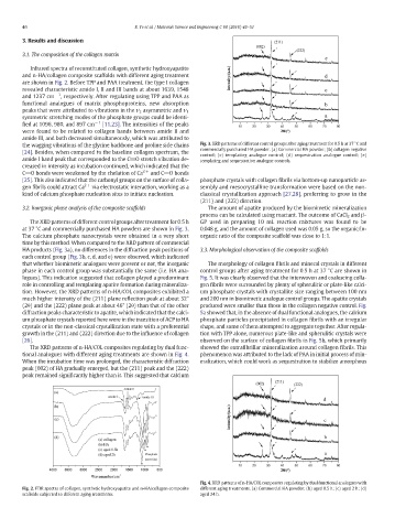
Infrared spectra of reconstituted collagen, synthetic hydroxyapatite
and n-HA/collagen composite scaffolds with different aging treatment
are shown in Fig. 2. Before TPP and PAA treatment, the type I collagen
revealed characteristic amide I, II and III bands at about 1639, 1548
and 1237 cm −1 , respectively. After regulating using TPP and PAA as
functional analogues of matrix phosphoproteins, new absorption
peaks that were attributed to vibrations in the ν 3 asymmetric and ν 1
symmetric stretching modes of the phosphate groups could be identi-
fied at 1096, 980, and 897 cm −1 [11,23]. The intensities of the peaks
were found to be related to collagen bands between amide II and
amide III, and both decreased simultaneously, which was attributed to
the wagging vibrations of the glycine backbone and proline side chains Fig. 3. XRD patterns of different control groups after aging treatment for 0.5 h at 37 °C and
commercially purchased HA powder. (a) Commercial HA powder; (b) collagen negative
[24]. Besides, when compared to the baseline collagen spectrum, the
control; (c) templating analogue control; (d) sequestration analogue control; (e)
amide I band peak that corresponded to the C_O stretch vibration de- templating and sequestration analogue controls.
creased in intensity as incubation continued, which indicated that the
C_O bonds were weakened by the chelation of Ca 2+ and C_Obonds
[25]. This also indicated that the carbonyl groups on the surface of colla- phosphate crystals with collagen fibrils via bottom-up nanoparticle as-
gen fibrils could attract Ca 2+ via electrostatic interaction, working as a sembly and mesocrystalline transformation were based on the non-
kind of calcium phosphate nucleation sites to initiate nucleation. classical crystallization approach [27,28],preferringtogrow inthe
(211) and (222) direction.
3.2. Inorganic phase analysis of the composite scaffolds The amount of apatite produced by the biomimetic mineralization
process can be calculated using reactant. The outcome of CaCl 2 and β-
The XRD patterns of different control groups after treatment for 0.5 h GP used in preparing 10 mL reaction mixtures was found to be
at 37 °C and commercially purchased HA powders are shown in Fig. 3. 0.048 g, and the amount of collagen used was 0.05 g, so the organic/in-
The calcium phosphate nanocrystals were obtained in a very short organic ratio of the composite scaffold was close to 1:1.
time by this method. When compared to the XRD pattern of commercial
HA products (Fig. 3a), no differences in the diffraction peak positions of 3.3. Morphological observation of the composite scaffolds
each control group (Fig. 3b, c, d, and e) were observed, which indicated
that whether biomimetic analogues were present or not, the inorganic The morphology of collagen fibrils and mineral crystals in different
phase in each control group was substantially the same (i.e. HA ana- control groups after aging treatment for 0.5 h at 37 °C are shown in
logues). This indication suggested that collagen played a predominant Fig. 5. It was clearly observed that the interwoven and coalescing colla-
role in controlling and templating apatite formation during mineraliza- gen fibrils were surrounded by plenty of spherulitic or plate-like calci-
tion. However, the XRD patterns of n-HA/COL composites exhibited a um phosphate crystals with crystallite size ranging between 100 nm
much higher intensity of the (211) plane reflection peak at about 32° and 200 nm in biomimetic analogue control groups. The apatite crystals
(2θ) and the (222) plane peak at about 46° (2θ) than that of the other produced were smaller than those in the collagen negative control. Fig.
diffraction peaks characteristic to apatite, which indicated that the calci- 5a showed that, in the absence of dual functional analogues, the calcium
um phosphate crystals reported here were in the transition of ACP to HA phosphate particles precipitated in collagen fibrils with an irregular
crystals or in the non-classical crystallization state with a preferential shape, and some of them attempted to aggregate together. After regula-
growth in the (211) and (222) direction due to the influence of collagen tion with TPP alone, numerous plate-like and spherulitic crystals were
[26]. observed on the surface of collagen fibrils in Fig. 5b, which primarily
The XRD patterns of n-HA/COL composites regulating by dual func- showed the extrafibrillar mineralization around collagen fibrils. This
tional analogues with different aging treatments are shown in Fig. 4. phenomenon was attributed to the lack of PAA in initial process of min-
When the incubation time was prolonged, the characteristic diffraction eralization, which could work as sequestration to stabilize amorphous
peak (002) of HA gradually emerged, but the (211) peak and the (222)
peak remained significantly higher than it. This suggested that calcium
Fig. 4. XRD patterns of n-HA/COL composites regulating by dual functional analogues with
Fig. 2. FTIR spectra of collagen, synthetic hydroxyapatite and n-HA/collagen composite different aging treatments. (a) Commercial HA powder; (b) aged 0.5 h; (c) aged 2 h; (d)
scaffolds subjected to different aging treatments. aged 24 h.