
Page 87 - Power of Stem Cells- arthritis and regeneration
P. 87
B. Ye et al. / Materials Science and Engineering C 68 (2016) 43–51 47
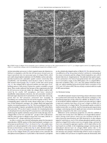
Fig. 5. FESEM images of collagen fibrils and mineral crystals in different control groups after aging treatment for 0.5 h at 37 °C. (a) Collagen negative control; (b) templating analogue
control; (c) sequestration analogue control; (d) templating and sequestration analogues control.
calcium phosphate precursors in their required nanoscale dimensions. on the cylindrically shaped surface of fibrils [30]. The selected area elec-
Without co-regulation with PAA, the ACP precursors formed were too tron diffraction of Fig. 6d was done to further confirm the relationship of
large to penetrate into the nanoscopic gaps of collagen fibrils, which the crystal orientation and the collagen fibrils longitudinal axis, which
just could only precipitate randomly over the fibrillar surface and subse- showed discrete arc-shaped patterns. This indicated that apatite report-
quently transform into spherulitic apatite crystals. Conversely, both ed was primarily in the form of nanopolycrystals, but some particles
extrafibrillar and interfibrillar mineralization could be observed in were in amorphous stages. In the SAED pattern, the arc-shaped diffrac-
PAA-treated control groups (Fig. 5c and d). In the PAA control group tions were ascribed to (002), (211), and (222) planes of HA from the in-
(Fig. 5c), the collagen fibrils appeared swollen and had vague cross- side out, which exhibited pronounced preferential orientation of the
banding patterns with plenty of plate-like apatite crystallites around (211) and (222) plane of HA. This was closely consistent with the results
them. These results indicated that because of the sequestration by PAA of XRD measurement.
for ACP precursors to become stable, the collagen fibrils could be infil-
trated by the smaller ACP nanophases, and hence introduced 3.4. Mechanical testing
intrafibrillar mineralization. However, the self-assembled swollen fi-
brils had vague characteristic D-periodicity at the same time, which sug- Unconfined uniaxial compression testing and porosity measurement
gested that the ACP precursors penetrated were incapable of were performed to determine the effect of biomimetic analogues on the
assembling and transforming into hierarchical apatite without TPP as mechanical properties of the n-HA/COL scaffolds. As shown in Table 1,
a templating agent. Unlike the results shown earlier here, in the pres- all mineralized scaffolds exhibited a reduced porosity and much higher
ence of dual biomimetic analogues, the collagen fibrils had apparently compressive modulus than those of the pure collagen scaffolds due to
regular cross-banding patterns (Fig. 5d). This was attributed to the hier- the mineralization of collagen fibrils. The generated apatite resulted in
archical assembly of apatite nanoplatelets directed by TPP within fibrils. reinforcement effect on scaffolds. And the templating and sequestration
More precisely, TPP was here found to simulate the templating function analogue control group showed significant improvement in mechanical
of phosphoproteins by attracting ACP nanoprecursors and initiating and properties compared to the other control groups.
modulating the apatite nucleation and growth within the gap zones of Differences in the mechanical properties of n-HA/COL scaffolds in
−
collagen fibril. As the free ionizable OH groups in TPP could interact this study were mostly due to the difference of collagen fibrils mineral-
with the amine groups in collagen to form ionic cross-links, with the re- ization among control groups, which was also consistent with the re-
−
maining free OH groups attracting ACP precursors. sults of porosity measurement and SEM observation. In the collagen
In order to obtain more detailed information about the biomimetic negative control group, most of the calcium phosphate particles precip-
mineralization, HRTEM analysis of the dual biomimetic analogues con- itated on collagen fibrils surface were in an irregular shape, and some of
trol group was performed in Fig. 6.Asshown in Fig. 6, the apatite nano- them even attempted to aggregate together, which might influence the
particles not only precipitated on the exterior collagen fibrils (Fig. 6b), load transfer in scaffolds. In the templating analogue control group, the
but also on the interior or at the ends (Fig. 6c and d, the short white ar- mineralized mineral layer on collagen fibrils surface due to the
rows). In Fig. 6 (c and d), the long white arrows indicated the longitudi- extrafibrillar mineralization resulted in the enhancement of compres-
nal axis of the collagen fibrils [29], and the black arrows indicated the sive modulus and strength. With the help of PAA to work as sequestra-
amorphous mineral phases or crystal defects that seemed to precipitate tion to stabilize ACP precursors in required nanoscale dimensions, both