
Page 90 - Power of Stem Cells- arthritis and regeneration
P. 90
50 B. Ye et al. / Materials Science and Engineering C 68 (2016) 43–51
Fig. 10. The 3D CT images 12 weeks after the operation.
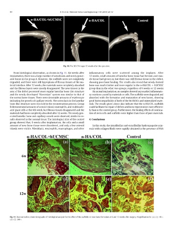
From histological observation, as shown in Fig. 11. Six weeks after inflammatory cells were scattered among the implants. After
implantation, there was a large number of osteoblasts and newly gener- 12 weeks, small amounts of lamellar bone tissue had formed, and mus-
ated bones in the group A. However, the scaffolds were not completely cle tissue had grown in, but there was still fibrous tissue in the defect,
degraded, and there were still hyperplasia of fibrous tissues at the ma- showing poor bone healing. The results also reveal that newly-formed
terial interface. After 12 weeks, the materials were completely absorbed bone was much thicker and more regular in the n-HA/COL + hUCMSC
and the fibrous layers were mostly disappeared. The new tissues in the group than in the other two groups, regardless of 6 weeks or 12 weeks.
area of the defect possessed more regular lamellar bone-like structure By animal implantation, no samples showed any marked inflammato-
and the newly developed “Haversian” system was similar to that of ry reactions caused by materials or cells. The scaffolds were degraded and
the normal bone tissues. There were detectable amounts of erythrocyte absorbed with the formation and maturation of new bones, showing
indicating the growth of capillary vessels. The osteoclasts in the lamellar good histocompatibility of both of the hUCMSCs and mineralized mate-
bone-like structure were involved in the reconstruction process. Group rials. The results given above also indicate that the n-HA/COL scaffolds
B demonstrated amounts of osteoid island, neutrophils, and multinucle- could facilitate the repair of defects and bone regeneration more efficient-
ated giant cells at the 6th week, but fibrous tissues disappeared and the ly than in the control group. Furthermore, the healing effects of combina-
materials had been completely absorbed after 12 weeks. The newly gen- tion of stem cells and scaffolds were higher than those of pure materials.
erated lamellar bone and capillary vessels were observed, similar to re-
sults observed in the normal tissue. The histological slice of the control 4. Conclusions
group showed that, 6 weeks after implantation, the cells and a small
amount of new bone tissue were disordered, and only a few osteoid In this study, the intrafibrillar and extrafibrillar hydroxyapatite min-
islands were visible. Fibroblasts, neutrophils, macrophages, and other erals with collagen fibrils were rapidly obtained in the presence of PAA
Fig. 11. Representative photomicrographs of histological analysis for the effect of the scaffolds on new bone formation at 6 and 12 weeks after surgery. Magnification for (a)–(c): 40×;
(d)–(f): 200×.