
Page 89 - Power of Stem Cells- arthritis and regeneration
P. 89
B. Ye et al. / Materials Science and Engineering C 68 (2016) 43–51 49
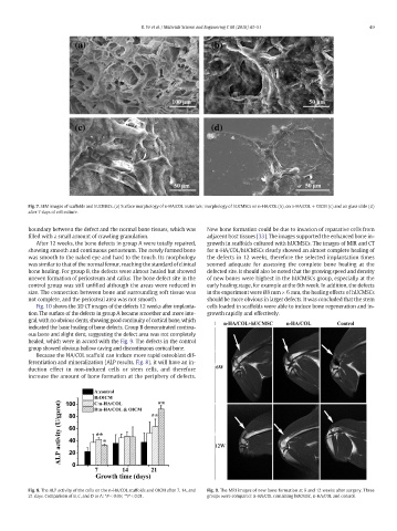
Fig. 7. SEM images of scaffolds and hUCMSCs. (a) Surface morphology of n-HA/COL materials; morphology of hUCMSCs on n-HA/COL (b), on n-HA/COL + OICM (c) and on glass slide (d)
after 7 days of cell culture.
boundary between the defect and the normal bone tissues, which was New bone formation could be due to invasion of reparative cells from
filled with a small amount of crawling granulation. adjacent host tissues [33]. The images supported the enhanced bone in-
After 12 weeks, the bone defects in group A were totally repaired, growth in scaffolds cultured with hUCMSCs. The images of MIR and CT
showing smooth and continuous periosteum. The newly formed bone for n-HA/COL/hUCMSCs clearly showed an almost complete healing of
was smooth to the naked eye and hard to the touch. Its morphology the defects in 12 weeks, therefore the selected implantation times
was similar to that of the normal femur, reaching the standard of clinical seemed adequate for assessing the complete bone healing at the
bone healing. For group B, the defects were almost healed but showed defected site. It should also be noted that the growing speed and density
uneven formation of periosteum and callus. The bone defect site in the of new bones were highest in the hUCMSCs group, especially at the
control group was still unfilled although the areas were reduced in early healing stage, for example at the 6th week. In addition, the defects
size. The connection between bone and surrounding soft tissue was in the experiment were Ø8 mm × 6 mm, the healing effects of hUCMSCs
not complete, and the periosteal area was not smooth. should be more obvious in larger defects. It was concluded that the stem
Fig. 10 shows the 3D CT images of the defects 12 weeks after implanta- cells loaded in scaffolds were able to induce bone regeneration and in-
tion. The surface of the defects in group A became smoother and more inte- growth rapidly and effectively.
gral, with no obvious dents, showing good continuity of cortical bone, which
indicated the basic healing of bone defects. Group B demonstrated continu-
ous bone and slight dent, suggesting the defect area was not completely
healed, which were in accord with the Fig. 9. The defects in the control
group showed obvious hollow caving and discontinuous cortical bone.
Because the HA/COL scaffold can induce more rapid osteoblast dif-
ferentiation and mineralization (ALP results, Fig. 8), it will have an in-
duction effect in non-induced cells or stem cells, and therefore
increase the amount of bone formation at the periphery of defects.
Fig. 8. The ALP activity of the cells on the n-HA/COL scaffolds and OICM after 7, 14, and Fig. 9. The MRI images of new bone formation at 6 and 12 weeks after surgery. Three
21 days. Comparison of B, C, and D to A: *P b 0.05; **P b 0.01. groups were compared: n-HA/COL containing hUCMSC, n-HA/COL and control.