Page 261 - Atlas of Small Animal CT and MRI
P. 261

Sella and Parasellar Region  251


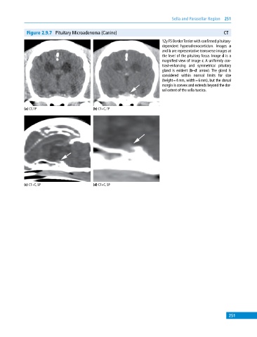
              Figure 2.9.7  Pituitary Microadenoma (Canine)                                               CT
                                                                             12y FS Border Terrier with confirmed pituitary‐
                                                                             dependent hyperadrenocorticism. Images  a
                                                                             and b are representative transverse images at
                                                                             the level of the pituitary fossa. Image d is a
                                                                             magnified view of image c. A uniformly con-
                                                                             trast‐enhancing and symmetrical pituitary
                                                                             gland is evident (b–d: arrow). The gland is
                                                                             considered within normal limits for size
                                                                             (height = 4 mm, width = 6 mm), but the dorsal
                                                                             margin is convex and extends beyond the dor-
                                                                             sal extent of the sella turcica.


            (a) CT, TP                       (b) CT+C, TP


            (c) CT+C, SP                     (d) CT+C, SP


                                                                                                             251
   256   257   258   259   260   261   262   263   264   265   266