
Page 265 - Atlas of Small Animal CT and MRI
P. 265
Sella and Parasellar Region 255
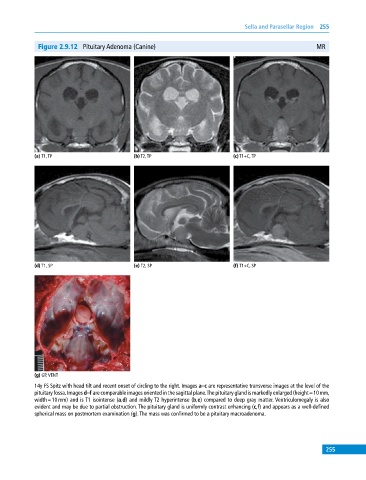
Figure 2.9.12 Pituitary Adenoma (Canine) MR
(a) T1, TP (b) T2, TP (c) T1+C, TP
(d) T1, SP (e) T2, SP (f) T1+C, SP
(g) GP, VENT
14y FS Spitz with head tilt and recent onset of circling to the right. Images a–c are representative transverse images at the level of the
pituitary fossa. Images d–f are comparable images oriented in the sagittal plane. The pituitary gland is markedly enlarged (height = 10 mm,
width = 10 mm) and is T1 isointense (a,d) and mildly T2 hyperintense (b,e) compared to deep gray matter. Ventriculomegaly is also
evident and may be due to partial obstruction. The pituitary gland is uniformly contrast enhancing (c,f) and appears as a well‐defined
spherical mass on postmortem examination (g). The mass was confirmed to be a pituitary macroadenoma.
255