
Page 267 - Atlas of Small Animal CT and MRI
P. 267
Sella and Parasellar Region 257
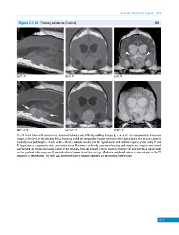
Figure 2.9.14 Pituitary Adenoma (Canine) MR
(a) T1, SP (b) T1, TP (c) T2, TP
(d) T1+C, SP (e) T1+C, TP (f) T2*, TP
11y FS Great Dane with recent‐onset abnormal behavior and difficulty walking. Images b, c, e, and f are representative transverse
images at the level of the pituitary fossa. Images a and d are comparable images oriented in the sagittal plane. The pituitary gland is
markedly enlarged (height = 17 mm, width = 30 mm), extends dorsally into the hypothalamic and thalamic regions, and is mildly T1 and
T2 hyperintense compared to deep gray matter (a–c). The mass is uniformly contrast enhancing, and margins are irregular and extend
well beyond the rostral and caudal extent of the pituitary fossa (d: arrows). Central mixed T2 intensity (c) and multifocal signal voids
on the gradient echo sequence (f) are indicative of parenchymal hemorrhage. Moderate peripheral edema is also evident on the T2
sequence (c: arrowheads). The mass was confirmed to be a pituitary adenoma on postmortem examination.
257