
Page 308 - Atlas of Small Animal CT and MRI
P. 308
298 Atlas of Small Animal CT and MRI
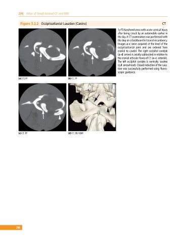
Figure 3.2.2 Occipitoatlantal Luxation (Canine) CT
1y FS Keeshond cross with acute cervical injury
after being struck by an automobile earlier in
the day. A CT examination was performed with
the dog on a backboard in lateral recumbency.
Images a–c were acquired at the level of the
occipitoatlantal joint and are ordered from
cranial to caudal. The right occipital condyle
(a–d: arrow) is axially subluxated in relation to
the cranial articular fovea of C1 (a–c: asterisk).
The left occipital condyle is ventrally luxated
(c,d: arrowhead). Closed reduction of the luxa-
tion was successfully performed using fluoro-
scopic guidance.
(a) CT, TP (b) CT, TP
(c) CT, TP (d) CT, 3D, VENT
298