
Page 311 - Atlas of Small Animal CT and MRI
P. 311
Traumatic and Vascular Disorders 301
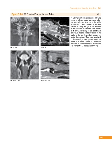
Figure 3.2.6 C2 Odontoid Process Fracture (Feline) MR
4y FS Bengal with generalized ataxia following
trauma of unknown cause. A displaced odon-
toid process fracture (a: arrow) and a mildly
displaced left C1 wing fracture (a: arrowhead)
are seen on survey radiographs. The odontoid
process fracture is also evident on MR images
(b–d: arrow). Instability of the atlantoaxial
joint results in spinal cord compression of the
cranial cervical spinal cord, best seen on the
sagittal images (b,d). There is an associated
focal region of T2 hyperintensity within the
ventral aspect of the spinal cord immediately
dorsal to the fractured odontoid process and
best seen on the T2 image (b: arrowhead).
(a) DX, VD (b) T2, SP
(c) SPGR+C, DP (d) SPGR+C, SP
301