
Page 316 - Atlas of Small Animal CT and MRI
P. 316
306 Atlas of Small Animal CT and MRI
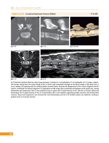
Figure 3.2.12 L3 Lamina Depression Fracture (Feline) CT & MR
(a) CT, TP (b) CT, SP (c) CT, 3D, OBL
(d) T2, TP (e) T2, SP
8y FS Domestic Shorthair bitten by a dog 2 days previously. Currently has a nonambulatory T3–L3 myelopathy. On CT images, a depres-
sion fracture arising from the cranial margin of the L3 lamina (a,b: arrowhead) impinges on the dorsal margin of the spinal cord. A
ventral oblique 3D rendering with the vertebral bodies removed further illustrates the displacement of the fracture fragment into the
canal (c: arrowhead). The fracture fragment is T2 hypointense on MR images (d,e: arrowhead) and impinges on the spinal cord, causing
deformation and compression. There is also marked increase in spinal cord T2 signal intensity at this level due to intrinsic edema and
hemorrhage. The T2 signal intensity of the L3–4 intervertebral disk is also reduced, suggesting possible traumatic intervertebral disk
extrusion. Spinal cord compression was relieved with a hemilaminectomy, and the L3–4 vertebral column was stabilized, resulting in
gradual return of neurologic function.
306