
Page 321 - Atlas of Small Animal CT and MRI
P. 321
Traumatic and Vascular Disorders 311
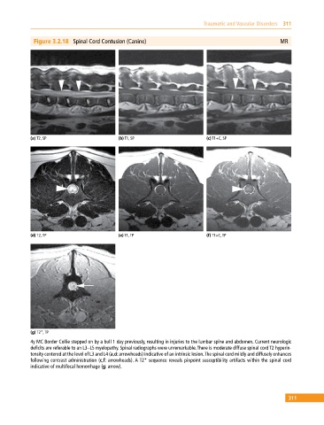
Figure 3.2.18 Spinal Cord Contusion (Canine) MR
(a) T2, SP (b) T1, SP (c) T1+C, SP
(d) T2, TP (e) T1, TP (f) T1+C, TP
(g) T2*, TP
4y MC Border Collie stepped on by a bull 1 day previously, resulting in injuries to the lumbar spine and abdomen. Current neurologic
deficits are referable to an L3–L5 myelopathy. Spinal radiographs were unremarkable. There is moderate diffuse spinal cord T2 hyperin-
tensity centered at the level of L3 and L4 (a,d: arrowheads) indicative of an intrinsic lesion. The spinal cord mildly and diffusely enhances
following contrast administration (c,f: arrowheads). A T2* sequence reveals pinpoint susceptibility artifacts within the spinal cord
indicative of multifocal hemorrhage (g: arrow).
311