
Page 322 - Atlas of Small Animal CT and MRI
P. 322
312 Atlas of Small Animal CT and MRI
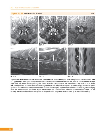
Figure 3.2.19 Hematomyelia (Canine) MR
(a) T1, TP (b) T2, TP (c) T2*, TP
(d) T1, SP (e) T2, SP (f) T2*, DP
12y FS Pit Bull Terrier with acute‐onset tetraparesis. The owners have administered aspirin twice weekly for chronic osteoarthrosis. There
is T2 hyperintensity of the spinal cord parenchyma and focal central canal dilation centered on C5 (b,e: arrow). Cord diameter is enlarged
at this level, indicative of intrinsic pathology, but there is no evidence of compression. The internal venous plexus also appears dilated
(a,b: arrowheads). T2* sequences document hemorrhage within the effected spinal cord segment, as evidenced by prominent susceptibil-
ity effect (c,f: arrowhead). Postmortem examination confirmed hematomyelia, myelomalacia, and subdural hemorrhage. An underlying
cause was not determined, and chronic aspirin administration was thought to have induced a spontaneous hemorrhage. The two
vertically oriented curvilinear lines superimposed on the spinal cord in image e are artifacts caused by an out‐of‐field microchip.
312