
Page 323 - Atlas of Small Animal CT and MRI
P. 323
Traumatic and Vascular Disorders 313
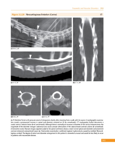
Figure 3.2.20 Fibrocartilaginous Embolism (Canine) CT
(a) DX+C, LAT
(c) CT+C, SP (b) CT+C, DP
(d) CT+C, TP (e) CT+C, TP
4y FS Yorkshire Terrier with peracute onset of tetraparesis shortly after returning from a walk with the owner. A myelographic examina-
tion reveals a pronounced increase in spinal cord diameter centered on C6 (a: arrowheads). CT myelography further documents a
caudal cervical intrinsic lesion (b–d: arrowheads). Although there is normally an increase in diameter of the cervical intumescence, the
magnitude of the diameter change is abnormal and causes annular attenuation of the subarachnoid contrast column (d: arrowhead).
A transverse cranial thoracic image acquired caudal to the spinal cord lesion shows a more normal spinal cord diameter and prominent
contrast‐enhanced subarachnoid space (e). Postmortem examination confirmed regional myelomalacia caused by multiple fibrocarti-
laginous emboli. The degree of spinal cord diameter enlargement in this patient is somewhat unusual and would be less likely to occur
in patients with recoverable disease.
313